2EDG | pdb_00002edg

Solution structure of the GCV_H domain from mouse glycine


Experimental Data Snapshot

  • Method: SOLUTION NMR
  • Conformers Calculated: 100 
  • Conformers Submitted: 20 
  • Selection Criteria: structures with the least restraint violations, target function 

wwPDB Validation 3D Report Full Report

Validation slider image for 2EDG

This is version 1.3 of the entry. See complete history

Literature

Solution structure of the GCV_H domain from mouse glycine

Endo, H.Yoshida, M.Hayashi, F.Yokoyama, S.

To be published.

Macromolecule Content 

  • Total Structure Weight: 14.19 kDa 
  • Atom Count: 994 
  • Modeled Residue Count: 130 
  • Deposited Residue Count: 130 
  • Unique protein chains: 1

Macromolecules

Find similar proteins by:|  3D Structure
Entity ID: 1
MoleculeChains  Sequence LengthOrganismDetailsImage
Glycine cleavage system H protein130Mus musculusMutation(s): 0 
Gene Names: Gcsh
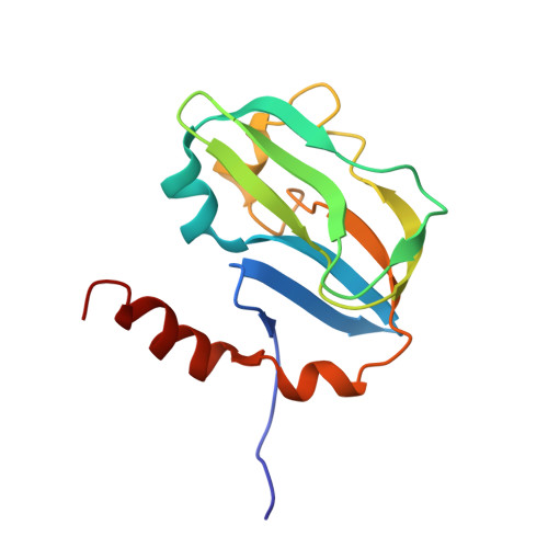
UniProt & NIH Common Fund Data Resources
Find proteins for Q91WK5 (Mus musculus)
Explore Q91WK5 
Go to UniProtKB:  Q91WK5
IMPC:  MGI:1915383
Entity Groups
Sequence Clusters30% Identity50% Identity70% Identity90% Identity95% Identity100% Identity
UniProt GroupQ91WK5
Sequence Annotations
Expand
Reference Sequence

Experimental Data & Validation

Experimental Data

  • Method: SOLUTION NMR
  • Conformers Calculated: 100 
  • Conformers Submitted: 20 
  • Selection Criteria: structures with the least restraint violations, target function 

Structure Validation

View Full Validation Report


Entry History 

Deposition Data

Revision History  (Full details and data files)

  • Version 1.0: 2007-08-14
    Type: Initial release
  • Version 1.1: 2011-07-13
    Changes: Version format compliance
  • Version 1.2: 2022-03-09
    Changes: Data collection, Database references, Derived calculations
  • Version 1.3: 2024-05-29
    Changes: Data collection