2E7S | pdb_00002e7s

Crystal structure of the yeast Sec2p GEF domain


Experimental Data Snapshot

  • Method: X-RAY DIFFRACTION
  • Resolution: 3.00 Å
  • R-Value Free: 
    0.285 (Depositor), 0.282 (DCC) 
  • R-Value Work: 
    0.229 (Depositor), 0.234 (DCC) 

wwPDB Validation 3D Report Full Report

Validation slider image for 2E7S

This is version 1.4 of the entry. See complete history

Literature

Asymmetric Coiled-Coil Structure with Guanine Nucleotide Exchange Activity

Sato, Y.Shirakawa, R.Horiuchi, H.Dohmae, N.Fukai, S.Nureki, O.

(2007) Structure 15: 245-252

  • DOI: https://doi.org/10.1016/j.str.2007.01.003
  • Primary Citation Related Structures: 
    2E7S

  • PubMed Abstract: 

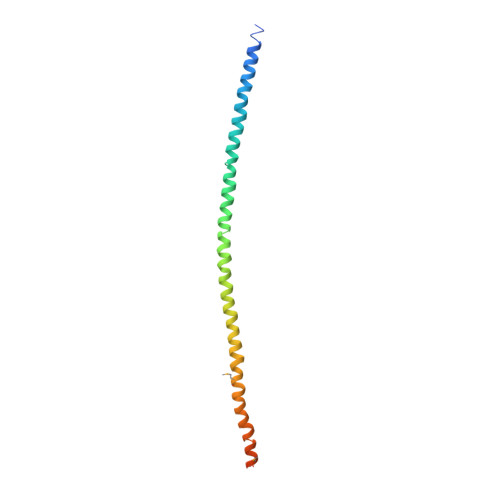
    Vesicular traffic during exocytosis is regulated by Rab GTPase, Sec4p in yeast, which is activated by a guanine nucleotide exchange factor (GEF) called Sec2p. The GEF activity is localized in the N-terminal 160 residues of Sec2p, which lacks sequence similarity with any other GEFs with known structures, and thereby the guanine nucleotide exchange mechanism by Sec2p remains unknown. Here, we report the crystal structure of the Sec2p GEF domain at 3.0 A resolution. The structure unexpectedly consists of a homodimeric, parallel coiled coil that extends over 180 A. Pull-down and guanine nucleotide exchange analyses on a series of deletion and point mutants of Sec2p unveiled the catalytic residues for its GEF activity as well as the Sec4p binding site, thus presenting a nucleotide exchange mechanism by a simple coiled coil. The present functional analyses allow us to build the Sec2p:Sec4p complex model, which explains the specificity for Rab GTPases by their respective GEF proteins.


  • Organizational Affiliation
    • Department of Biological Information, Graduate School of Bioscience and Biotechnology, Tokyo Institute of Technology, 4259 Nagatsuta-cho, Yokohama-shi, Kanagawa, Japan.

Macromolecule Content 

  • Total Structure Weight: 315.61 kDa 
  • Atom Count: 19,270 
  • Modeled Residue Count: 2,313 
  • Deposited Residue Count: 2,700 
  • Unique protein chains: 1

Macromolecules

Find similar proteins by:|  3D Structure
Entity ID: 1
MoleculeChains  Sequence LengthOrganismDetailsImage
Rab guanine nucleotide exchange factor SEC2
A, B, C, D, E
A, B, C, D, E, F, G, H, I, J, K, L, M, N, O, P, Q, R, S, T
135Saccharomyces cerevisiaeMutation(s): 1 
UniProt
Find proteins for P17065 (Saccharomyces cerevisiae (strain ATCC 204508 / S288c))
Explore P17065 
Go to UniProtKB:  P17065
Entity Groups
Sequence Clusters30% Identity50% Identity70% Identity90% Identity95% Identity100% Identity
UniProt GroupP17065
Sequence Annotations
Expand
Reference Sequence

Small Molecules

Modified Residues  1 Unique
IDChains TypeFormula2D DiagramParent
MSE
Query on MSE
A, B, C, D, E
A, B, C, D, E, F, G, H, I, J, K, L, M, N, O, P, Q, R, S, T
L-PEPTIDE LINKINGC5 H11 N O2 SeMET

Experimental Data & Validation

Experimental Data

  • Method: X-RAY DIFFRACTION
  • Resolution: 3.00 Å
  • R-Value Free:  0.285 (Depositor), 0.282 (DCC) 
  • R-Value Work:  0.229 (Depositor), 0.234 (DCC) 
Space Group: P 1 21 1
Unit Cell:
Length ( Å )Angle ( ˚ )
a = 101.937α = 90
b = 176.569β = 90
c = 181.453γ = 90
Software Package:
Software NamePurpose
ADSCdata collection
SHARPphasing
CNSrefinement
DENZOdata reduction
SCALEPACKdata scaling

Structure Validation

View Full Validation Report


Entry History 

Deposition Data

Revision History  (Full details and data files)

  • Version 1.0: 2007-02-27
    Type: Initial release
  • Version 1.1: 2008-04-30
    Changes: Version format compliance
  • Version 1.2: 2011-07-13
    Changes: Version format compliance
  • Version 1.3: 2021-11-10
    Changes: Database references, Derived calculations
  • Version 1.4: 2024-10-30
    Changes: Data collection, Structure summary