2E7D | pdb_00002e7d

Crystal structure of a NEAT domain from Staphylococcus aureus


Experimental Data Snapshot

  • Method: X-RAY DIFFRACTION
  • Resolution: 2.20 Å
  • R-Value Free: 
    0.265 (Depositor), 0.262 (DCC) 
  • R-Value Work: 
    0.240 (Depositor), 0.240 (DCC) 
  • R-Value Observed: 
    0.240 (Depositor) 

wwPDB Validation 3D Report Full Report

Validation slider image for 2E7D

This is version 1.2 of the entry. See complete history

Literature

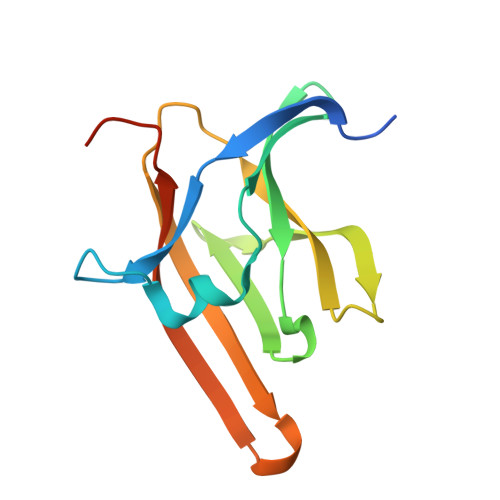
Structural basis for multimeric heme complexation through a specific protein-heme interaction: the case of the third neat domain of IsdH from Staphylococcus aureus

Watanabe, M.Tanaka, Y.Suenaga, A.Kuroda, M.Yao, M.Watanabe, N.Arisaka, F.Ohta, T.Tanaka, I.Tsumoto, K.

(2008) J Biological Chem 283: 28649-28659

  • DOI: https://doi.org/10.1074/jbc.M803383200
  • Primary Citation Related Structures: 
    2E7D, 2Z6F

  • PubMed Abstract: 

    To elucidate the heme acquisition system in pathogenic bacteria, we investigated the heme-binding properties of the third NEAT domain of IsdH (IsdH-NEAT3), a receptor for heme located on the surfaces of pathogenic bacterial cells, by using x-ray crystallography, isothermal titration calorimetry, examination of absorbance spectra, mutation analysis, size-exclusion chromatography, and analytical ultracentrifugation. We found the following: 1) IsdH-NEAT3 can bind with multiple heme molecules by two modes; 2) heme was bound at the surface of IsdH-NEAT3; 3) candidate residues proposed from the crystal structure were not essential for binding with heme; and 4) IsdH-NEAT3 was associated into a multimeric heme complex by the addition of excess heme. From these observations, we propose a heme-binding mechanism for IsdH-NEAT3 that involves multimerization and discuss the biological importance of this mechanism.


  • Organizational Affiliation
    • Department of Medical Genome Sciences, Graduate School of Frontier Sciences, the University of Tokyo, Tokyo 277-8562, Japan.

Macromolecule Content 

  • Total Structure Weight: 30.24 kDa 
  • Atom Count: 1,972 
  • Modeled Residue Count: 228 
  • Deposited Residue Count: 262 
  • Unique protein chains: 1

Macromolecules

Find similar proteins by:|  3D Structure
Entity ID: 1
MoleculeChains  Sequence LengthOrganismDetailsImage
Hypothetical protein IsdH
A, B
131Staphylococcus aureusMutation(s): 0 
Gene Names: IsdH
UniProt
Find proteins for Q931P4 (Staphylococcus aureus (strain Mu50 / ATCC 700699))
Explore Q931P4 
Go to UniProtKB:  Q931P4
Entity Groups
Sequence Clusters30% Identity50% Identity70% Identity90% Identity95% Identity100% Identity
UniProt GroupQ931P4
Sequence Annotations
Expand
Reference Sequence

Experimental Data & Validation

Experimental Data

  • Method: X-RAY DIFFRACTION
  • Resolution: 2.20 Å
  • R-Value Free:  0.265 (Depositor), 0.262 (DCC) 
  • R-Value Work:  0.240 (Depositor), 0.240 (DCC) 
  • R-Value Observed: 0.240 (Depositor) 
Space Group: H 3 2
Unit Cell:
Length ( Å )Angle ( ˚ )
a = 124.247α = 90
b = 124.247β = 90
c = 119.655γ = 120
Software Package:
Software NamePurpose
CNSrefinement
ADSCdata collection
HKL-2000data reduction
HKL-2000data scaling
SOLVEphasing

Structure Validation

View Full Validation Report


Entry History 

Deposition Data

Revision History  (Full details and data files)

  • Version 1.0: 2008-01-22
    Type: Initial release
  • Version 1.1: 2011-07-13
    Changes: Non-polymer description, Version format compliance
  • Version 1.2: 2024-03-13
    Changes: Data collection, Database references, Derived calculations