2E28

Crystal structure analysis of pyruvate kinase from Bacillus stearothermophilus


Experimental Data Snapshot

  • Method: X-RAY DIFFRACTION
  • Resolution: 2.40 Å
  • R-Value Free: 0.294 
  • R-Value Work: 0.226 

Starting Model: experimental
View more details

wwPDB Validation   3D Report Full Report


This is version 1.3 of the entry. See complete history


Literature

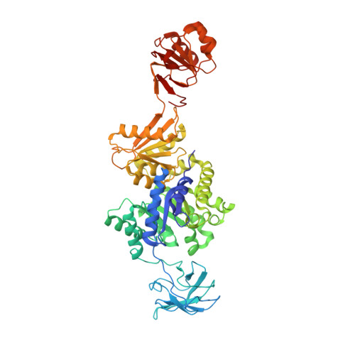
Crystal structure of pyruvate kinase from Geobacillus stearothermophilus.

Suzuki, K.Ito, S.Shimizu-Ibuka, A.Sakai, H.

(2008) J Biochem 144: 305-312

  • DOI: https://doi.org/10.1093/jb/mvn069
  • Primary Citation of Related Structures:  
    2E28

  • PubMed Abstract: 

    The pyruvate kinase (PK) from a moderate thermophile, Geobacillus stearothermophilus, is an allosteric enzyme activated by AMP and ribose 5-phosphate but not fructose 1, 6-bisphosphate (FBP), which is a common activator of PKs. It has an extra C-terminal sequence (ECTS), which contains a highly conserved phosphoenolpyruvate (PEP) binding motif, but its function and structure remain unclear. To elucidate the structural characteristics of the effector-binding site and the ECTS, the crystal structure of the C9S/C268S mutant of the enzyme was determined at 2.4 A resolution. The crystal belonged to space group P6(2)22, with unit cell parameters a, b = 145.97 A, c = 118.03 A. The enzyme was a homotetramer and its overall domain structure was similar to the previously solved structures except that the ECTS formed a new domain (C' domain). The structure of the C' domain closely resembled that of the PEP binding domain of maize pyruvate phosphate dikinase. A sulphate ion was found in a pocket in the effector-binding C domain. This site corresponds to the 6-phosphate group-binding site in yeast PK bound FBP and seems to be the effector-binding site. Through comparison of the structure of the putative effector-binding site to that of the FBP binding site of the yeast enzyme, the structural basis of the effector specificity of the G. stearothermophilus PK is discussed.


  • Organizational Affiliation

    Department of Food and Nutritional Sciences, University of Shizuoka, Yada 52-1, Shizuoka 422-8526, Japan.


Macromolecules
Find similar proteins by:  (by identity cutoff)  |  3D Structure
Entity ID: 1
MoleculeChains Sequence LengthOrganismDetailsImage
Pyruvate kinase587Geobacillus stearothermophilusMutation(s): 2 
EC: 2.7.1.40
UniProt
Find proteins for Q02499 (Geobacillus stearothermophilus)
Explore Q02499 
Go to UniProtKB:  Q02499
Entity Groups  
Sequence Clusters30% Identity50% Identity70% Identity90% Identity95% Identity100% Identity
UniProt GroupQ02499
Sequence Annotations
Expand
  • Reference Sequence
Small Molecules
Ligands 1 Unique
IDChains Name / Formula / InChI Key2D Diagram3D Interactions
SO4
Query on SO4

Download Ideal Coordinates CCD File 
B [auth A]SULFATE ION
O4 S
QAOWNCQODCNURD-UHFFFAOYSA-L
Experimental Data & Validation

Experimental Data

  • Method: X-RAY DIFFRACTION
  • Resolution: 2.40 Å
  • R-Value Free: 0.294 
  • R-Value Work: 0.226 
  • Space Group: P 62 2 2
Unit Cell:
Length ( Å )Angle ( ˚ )
a = 145.97α = 90
b = 145.97β = 90
c = 118.03γ = 120
Software Package:
Software NamePurpose
HKL-2000data collection
MOLREPphasing
CNSrefinement
HKL-2000data reduction
HKL-2000data scaling

Structure Validation

View Full Validation Report


Entry History 

Deposition Data

  • Released Date: 2008-02-05 
  • Deposition Author(s): Suzuki, K.

Revision History  (Full details and data files)

  • Version 1.0: 2008-02-05
    Type: Initial release
  • Version 1.1: 2011-07-13
    Changes: Version format compliance
  • Version 1.2: 2021-11-10
    Changes: Database references, Derived calculations
  • Version 1.3: 2023-10-25
    Changes: Data collection, Refinement description