2E1P | pdb_00002e1p

Crystal structure of pro-Tk-subtilisin


Experimental Data Snapshot

  • Method: X-RAY DIFFRACTION
  • Resolution: 2.30 Å
  • R-Value Free: 
    0.243 (Depositor), 0.243 (DCC) 
  • R-Value Work: 
    0.192 (Depositor), 0.192 (DCC) 
  • R-Value Observed: 
    0.192 (Depositor) 

Starting Model: experimental
View more details

wwPDB Validation 3D Report Full Report

Validation slider image for 2E1P

This is version 1.5 of the entry. See complete history

Literature

Crystal structure of unautoprocessed precursor of subtilisin from a hyperthermophilic archaeon: evidence for Ca2+-induced folding

Tanaka, S.Saito, K.Chon, H.Matsumura, H.Koga, Y.Takano, K.Kanaya, S.

(2007) J Biological Chem 282: 8246-8255

  • DOI: https://doi.org/10.1074/jbc.M610137200
  • Primary Citation Related Structures: 
    2E1P

  • PubMed Abstract: 

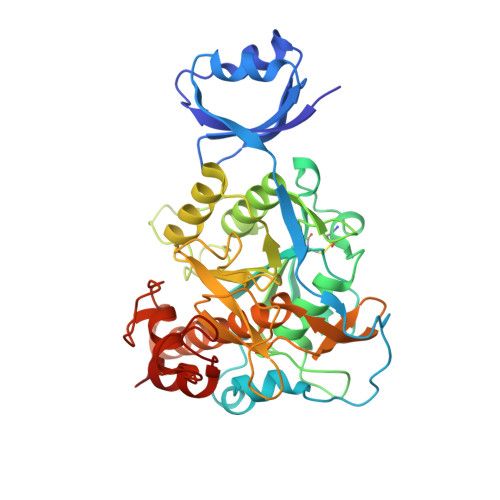
    The crystal structure of an active site mutant of pro-Tk-subtilisin (pro-S324A) from the hyperthermophilic archaeon Thermococcus kodakaraensis was determined at 2.3 A resolution. The overall structure of this protein is similar to those of bacterial subtilisin-propeptide complexes, except that the peptide bond linking the propeptide and mature domain contacts with the active site, and the mature domain contains six Ca2+ binding sites. The Ca-1 site is conserved in bacterial subtilisins but is formed prior to autoprocessing, unlike the corresponding sites of bacterial subtilisins. All other Ca2+-binding sites are unique in the pro-S324A structure and are located at the surface loops. Four of them apparently contribute to the stability of the central alphabetaalpha substructure of the mature domain. The CD spectra, 1-anilino-8-naphthalenesulfonic acid fluorescence spectra, and sensitivities to chymotryptic digestion of this protein indicate that the conformation of pro-S324A is changed from an unstable molten globule-like structure to a stable native one upon Ca2+ binding. Another active site mutant, pro-S324C, was shown to be autoprocessed to form a propeptide-mature domain complex in the presence of Ca2+. The CD spectra of this protein indicate that the structure of pro-S324C is changed upon Ca2+ binding like pro-S324A but is not seriously changed upon subsequent autoprocessing. These results suggest that the maturation process of Tk-subtilisin is different from that of bacterial subtilisins in terms of the requirement of Ca2+ for folding of the mature domain and completion of the folding process prior to autoprocessing.


  • Organizational Affiliation
    • Department of Material and Life Science, Graduate School of Engineering, Osaka University, 2-1 Yamadaoka, Suita, Osaka 565-0871, Japan.

Macromolecule Content 

  • Total Structure Weight: 41.63 kDa 
  • Atom Count: 3,170 
  • Modeled Residue Count: 395 
  • Deposited Residue Count: 398 
  • Unique protein chains: 1

Macromolecules

Find similar proteins by:|  3D Structure
Entity ID: 1
MoleculeChains  Sequence LengthOrganismDetailsImage
Tk-subtilisin398Thermococcus kodakarensis KOD1Mutation(s): 1 
EC: 3.4.21.62 (PDB Primary Data), 3.4.21 (UniProt)
UniProt
Find proteins for P58502 (Thermococcus kodakarensis (strain ATCC BAA-918 / JCM 12380 / KOD1))
Explore P58502 
Go to UniProtKB:  P58502
Entity Groups
Sequence Clusters30% Identity50% Identity70% Identity90% Identity95% Identity100% Identity
UniProt GroupP58502
Sequence Annotations
Expand
Reference Sequence

Experimental Data & Validation

Experimental Data

  • Method: X-RAY DIFFRACTION
  • Resolution: 2.30 Å
  • R-Value Free:  0.243 (Depositor), 0.243 (DCC) 
  • R-Value Work:  0.192 (Depositor), 0.192 (DCC) 
  • R-Value Observed: 0.192 (Depositor) 
Space Group: I 2 2 2
Unit Cell:
Length ( Å )Angle ( ˚ )
a = 92.69α = 90
b = 121.78β = 90
c = 77.53γ = 90
Software Package:
Software NamePurpose
CNSrefinement
HKL-2000data collection
HKL-2000data reduction
HKL-2000data scaling
MOLREPphasing

Structure Validation

View Full Validation Report


Entry History 

Deposition Data

Revision History  (Full details and data files)

  • Version 1.0: 2007-01-16
    Type: Initial release
  • Version 1.1: 2008-04-30
    Changes: Version format compliance
  • Version 1.2: 2011-07-13
    Changes: Source and taxonomy, Version format compliance
  • Version 1.3: 2021-11-10
    Changes: Database references, Derived calculations
  • Version 1.4: 2023-10-25
    Changes: Data collection, Refinement description
  • Version 1.5: 2024-10-23
    Changes: Structure summary