2DXB | pdb_00002dxb

Recombinant thiocyanate hydrolase comprising partially-modified cobalt centers


Experimental Data Snapshot

  • Method: X-RAY DIFFRACTION
  • Resolution: 2.25 Å
  • R-Value Free: 
    0.198 (Depositor), 0.167 (DCC) 
  • R-Value Work: 
    0.171 (Depositor), 0.167 (DCC) 
  • R-Value Observed: 
    0.171 (Depositor) 

Starting Model: experimental
View more details

wwPDB Validation 3D Report Full Report

Validation slider image for 2DXB

This is version 1.4 of the entry. See complete history

Literature

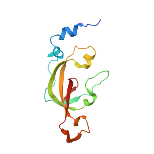
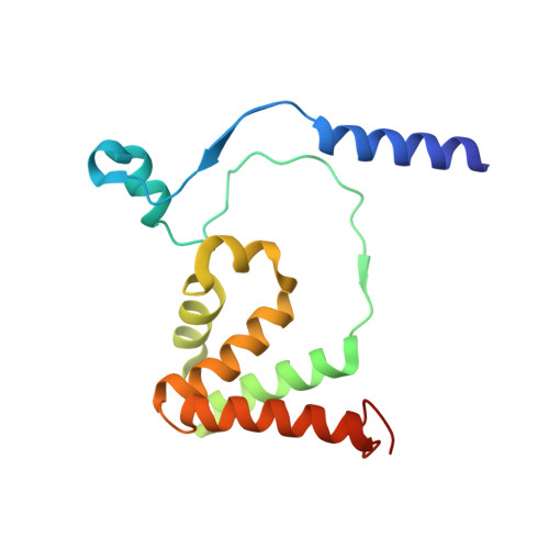
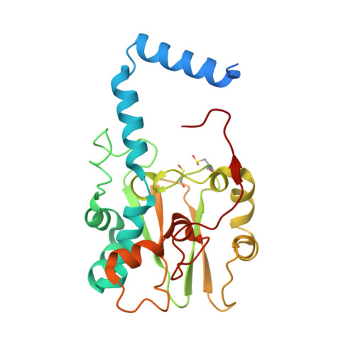
Structural Basis for Catalytic Activation of Thiocyanate Hydrolase Involving Metal-Ligated Cysteine Modification

Arakawa, T.Kawano, Y.Katayama, Y.Nakayama, H.Dohmae, N.Yohda, M.Odaka, M.

(2009) J Am Chem Soc 131: 14838-14843

  • DOI: https://doi.org/10.1021/ja903979s
  • Primary Citation Related Structures: 
    2DXB, 2DXC, 2ZZD

  • PubMed Abstract: 

    Thiocyanate hydrolase (SCNase) is a member of a family of nitrile hydratase proteins, each of which contains a unique noncorrin cobalt center with two post-translationally modified cysteine ligands, cysteine-sulfenic acid or -sulfenate (Cys-SO(H)), and cysteine-sulfininate (Cys-SO(2)(-)), respectively. We have found that a partially matured recombinant SCNase was activated during storage. The crystal structures of SCNase before and after storage demonstrated that Cys-SO(2)(-) modification of gammaCys131 proceeded to completion prior to storage, while Cys-SO(H) modification of gammaCys133 occurred during storage. SCNase activity was suppressed when gammaCys133 was further oxidized to Cys-SO(2)(-). The correlation between the catalytic activity and the extent of the gammaCys133 modification indicates that the cysteine sulfenic acid modification of gammaCys133 is of primary importance in determining the activity of SCNase.


  • Organizational Affiliation
    • Department of Biotechnology and Life Science, Graduate School of Technology, Tokyo University of Agriculture and Technology, Koganei, Tokyo 184-8588, Japan.

Macromolecule Content 

  • Total Structure Weight: 482.77 kDa 
  • Atom Count: 34,391 
  • Modeled Residue Count: 3,908 
  • Deposited Residue Count: 4,208 
  • Unique protein chains: 3

Macromolecules

Find similar proteins by:|  3D Structure
Entity ID: 1
MoleculeChains  Sequence LengthOrganismDetailsImage
Thiocyanate hydrolase subunit alpha
A, D, G, J, M
A, D, G, J, M, P, S, V
126Thiobacillus thioparusMutation(s): 0 
EC: 3.5.5.8
UniProt
Find proteins for O66187 (Thiobacillus thioparus)
Explore O66187 
Go to UniProtKB:  O66187
Entity Groups
Sequence Clusters30% Identity50% Identity70% Identity90% Identity95% Identity100% Identity
UniProt GroupO66187
Sequence Annotations
Expand
Reference Sequence
Find similar proteins by:|  3D Structure
Entity ID: 2
MoleculeChains  Sequence LengthOrganismDetailsImage
Thiocyanate hydrolase subunit beta
B, E, H, K, N
B, E, H, K, N, Q, T, W
157Thiobacillus thioparusMutation(s): 0 
EC: 3.5.5.8
UniProt
Find proteins for O66186 (Thiobacillus thioparus)
Explore O66186 
Go to UniProtKB:  O66186
Entity Groups
Sequence Clusters30% Identity50% Identity70% Identity90% Identity95% Identity100% Identity
UniProt GroupO66186
Sequence Annotations
Expand
Reference Sequence
Find similar proteins by:|  3D Structure
Entity ID: 3
MoleculeChains  Sequence LengthOrganismDetailsImage
Thiocyanate hydrolase subunit gamma
C, F, I, L, O
C, F, I, L, O, R, U, X
243Thiobacillus thioparusMutation(s): 0 
EC: 3.5.5.8
UniProt
Find proteins for O66188 (Thiobacillus thioparus)
Explore O66188 
Go to UniProtKB:  O66188
Entity Groups
Sequence Clusters30% Identity50% Identity70% Identity90% Identity95% Identity100% Identity
UniProt GroupO66188
Sequence Annotations
Expand
Reference Sequence

Small Molecules

Ligands 2 Unique
IDChains Name / Formula / InChI Key2D Diagram3D Interactions
PO4

Query on PO4


Download:Ideal Coordinates CCD File
BA [auth F]
DA [auth I]
FA [auth L]
HA [auth O]
JA [auth R]
BA [auth F],
DA [auth I],
FA [auth L],
HA [auth O],
JA [auth R],
LA [auth U],
NA [auth X],
Z [auth C]
PHOSPHATE ION
O4 P
NBIIXXVUZAFLBC-UHFFFAOYSA-K
3CO

Query on 3CO


Download:Ideal Coordinates CCD File
AA [auth F]
CA [auth I]
EA [auth L]
GA [auth O]
IA [auth R]
AA [auth F],
CA [auth I],
EA [auth L],
GA [auth O],
IA [auth R],
KA [auth U],
MA [auth X],
Y [auth C]
COBALT (III) ION
Co
JAWGVVJVYSANRY-UHFFFAOYSA-N
Modified Residues  2 Unique
IDChains TypeFormula2D DiagramParent
CSD
Query on CSD
C, F, I, L, O
C, F, I, L, O, R, U, X
L-PEPTIDE LINKINGC3 H7 N O4 SCYS
CSO
Query on CSO
C, F, I, L, O
C, F, I, L, O, R, U, X
L-PEPTIDE LINKINGC3 H7 N O3 SCYS

Experimental Data & Validation

Experimental Data

  • Method: X-RAY DIFFRACTION
  • Resolution: 2.25 Å
  • R-Value Free:  0.198 (Depositor), 0.167 (DCC) 
  • R-Value Work:  0.171 (Depositor), 0.167 (DCC) 
  • R-Value Observed: 0.171 (Depositor) 
Space Group: P 21 21 21
Unit Cell:
Length ( Å )Angle ( ˚ )
a = 114.874α = 90
b = 245.141β = 90
c = 244.653γ = 90
Software Package:
Software NamePurpose
CNSrefinement
HKL-2000data collection
HKL-2000data reduction
HKL-2000data scaling
MOLREPphasing

Structure Validation

View Full Validation Report


Entry History 

Deposition Data

Revision History  (Full details and data files)

  • Version 1.0: 2007-08-28
    Type: Initial release
  • Version 1.1: 2011-07-13
    Changes: Derived calculations, Version format compliance
  • Version 1.2: 2023-10-25
    Changes: Data collection, Database references, Derived calculations, Refinement description
  • Version 1.3: 2023-11-15
    Changes: Data collection
  • Version 1.4: 2024-10-23
    Changes: Structure summary