2DGN | pdb_00002dgn

Mouse Muscle Adenylosuccinate Synthetase partially ligated complex with GTP, 2'-deoxy-IMP


Experimental Data Snapshot

  • Method: X-RAY DIFFRACTION
  • Resolution: 2.40 Å
  • R-Value Free: 
    0.262 (Depositor), 0.257 (DCC) 
  • R-Value Work: 
    0.198 (Depositor), 0.195 (DCC) 

wwPDB Validation 3D Report Full Report

Validation slider image for 2DGN

Ligand Structure Quality Assessment 


This is version 1.3 of the entry. See complete history

Literature

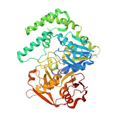
Cavitation as a mechanism of substrate discrimination by adenylosuccinate synthetases.

Iancu, C.V.Zhou, Y.Borza, T.Fromm, H.J.Honzatko, R.B.

(2006) Biochemistry 45: 11703-11711

  • DOI: https://doi.org/10.1021/bi0607498
  • Primary Citation Related Structures: 
    2DGN, 2GCQ

  • PubMed Abstract: 

    Adenylosuccinate synthetase catalyzes the first committed step in the de novo biosynthesis of AMP, coupling L-aspartate and IMP to form adenylosuccinate. Km values of IMP and 2'-deoxy-IMP are nearly identical with each substrate supporting comparable maximal velocities. Nonetheless, the Km value for L-aspartate and the Ki value for hadacidin (a competitive inhibitor with respect to L-aspartate) are 29-57-fold lower in the presence of IMP than in the presence of 2'-deoxy-IMP. Crystal structures of the synthetase ligated with hadacidin, GDP, and either 6-phosphoryl-IMP or 2'-deoxy-6-phosphoryl-IMP are identical except for the presence of a cavity normally occupied by the 2'-hydroxyl group of IMP. In the presence of 6-phosphoryl-IMP and GDP (hadacidin absent), the L-aspartate pocket can retain its fully ligated conformation, forming hydrogen bonds between the 2'-hydroxyl group of IMP and sequence-invariant residues. In the presence of 2'-deoxy-6-phosphoryl-IMP and GDP, however, the L-aspartate pocket is poorly ordered. The absence of the 2'-hydroxyl group of the deoxyribonucleotide may destabilize binding of the ligand to the L-aspartate pocket by disrupting hydrogen bonds that maintain a favorable protein conformation and by the introduction of a cavity into the fully ligated active site. At an approximate energy cost of 2.2 kcal/mol, the unfavorable thermodynamics of cavity formation may be the major factor in destabilizing ligands at the L-aspartate pocket.


  • Organizational Affiliation
    • Department of Biochemistry, Biophysics, and Molecular Biology, Iowa State University, Ames, Iowa 50011, USA.

Macromolecule Content 

  • Total Structure Weight: 51.2 kDa 
  • Atom Count: 3,535 
  • Modeled Residue Count: 431 
  • Deposited Residue Count: 457 
  • Unique protein chains: 1

Macromolecules

Find similar proteins by:|  3D Structure
Entity ID: 1
MoleculeChains  Sequence LengthOrganismDetailsImage
Adenylosuccinate synthetase isozyme 1457Mus musculusMutation(s): 0 
EC: 6.3.4.4
UniProt
Find proteins for P28650 (Mus musculus)
Explore P28650 
Go to UniProtKB:  P28650
Entity Groups
Sequence Clusters30% Identity50% Identity70% Identity90% Identity95% Identity100% Identity
UniProt GroupP28650
Sequence Annotations
Expand
Reference Sequence

Small Molecules

Ligands 3 Unique
IDChains Name / Formula / InChI Key2D Diagram3D Interactions
GDP

Query on GDP


Download:Ideal Coordinates CCD File
D [auth A]GUANOSINE-5'-DIPHOSPHATE
C10 H15 N5 O11 P2
QGWNDRXFNXRZMB-UUOKFMHZSA-N
DOI

Query on DOI


Download:Ideal Coordinates CCD File
C [auth A]9-(2-DEOXY-5-O-PHOSPHONO-BETA-D-ERYTHRO-PENTOFURANOSYL)-6-(PHOSPHONOOXY)-9H-PURINE
C10 H14 N4 O10 P2
GEECATYOCBMMIR-RRKCRQDMSA-N
MG

Query on MG


Download:Ideal Coordinates CCD File
B [auth A]MAGNESIUM ION
Mg
JLVVSXFLKOJNIY-UHFFFAOYSA-N

Experimental Data & Validation

Experimental Data

  • Method: X-RAY DIFFRACTION
  • Resolution: 2.40 Å
  • R-Value Free:  0.262 (Depositor), 0.257 (DCC) 
  • R-Value Work:  0.198 (Depositor), 0.195 (DCC) 
Space Group: P 43 21 2
Unit Cell:
Length ( Å )Angle ( ˚ )
a = 70.112α = 90
b = 70.112β = 90
c = 199.033γ = 90
Software Package:
Software NamePurpose
CrystalCleardata collection
TRUNCATEdata reduction
AMoREphasing
CNSrefinement
CrystalCleardata reduction
CCP4data scaling

Structure Validation

View Full Validation Report


Ligand Structure Quality Assessment 


Entry History 

Deposition Data

Revision History  (Full details and data files)

  • Version 1.0: 2006-09-15
    Type: Initial release
  • Version 1.1: 2008-04-30
    Changes: Version format compliance
  • Version 1.2: 2011-07-13
    Changes: Derived calculations, Version format compliance
  • Version 1.3: 2024-03-13
    Changes: Data collection, Database references, Derived calculations