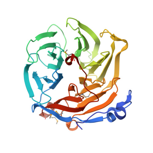
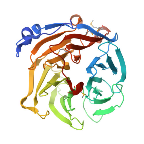
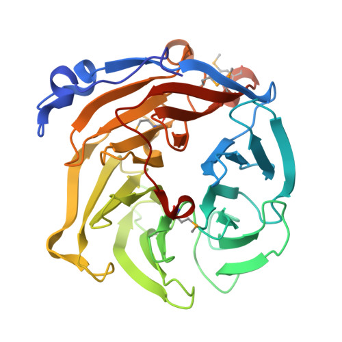
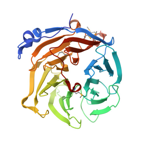
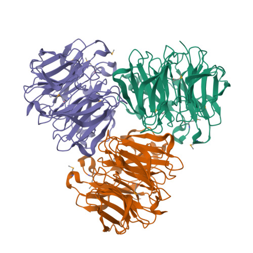
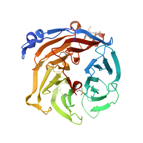
2DG0

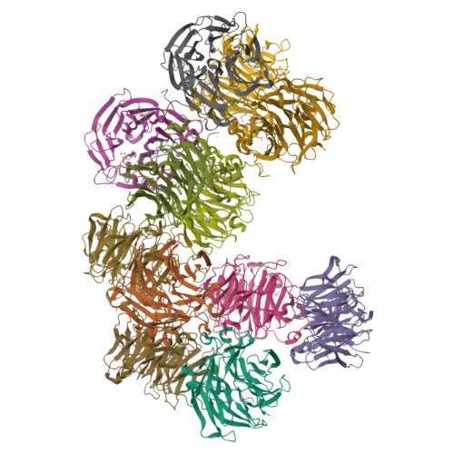
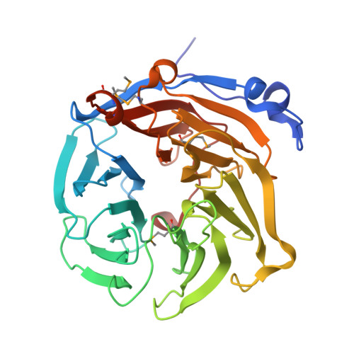
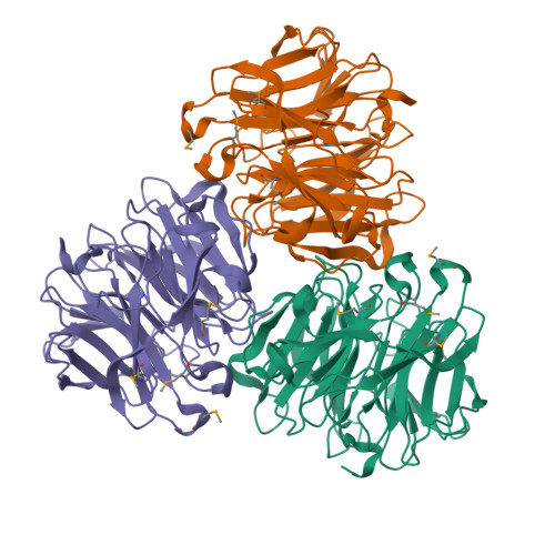
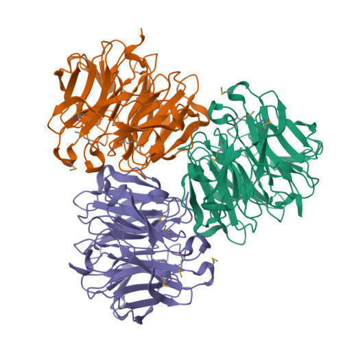
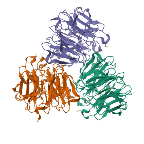
Crystal structure of Drp35, a 35kDa drug responsive protein from Staphylococcus aureus


Experimental Data Snapshot

  • Method: X-RAY DIFFRACTION
  • Resolution: 2.40 Å
  • R-Value Free: 0.222 
  • R-Value Work: 0.200 
  • R-Value Observed: 0.200 

wwPDB Validation   3D Report Full Report


This is version 1.3 of the entry. See complete history


Literature

Structural and Mutational Analyses of Drp35 from Staphylococcus aureus: A POSSIBLE MECHANISM FOR ITS LACTONASE ACTIVITY

Tanaka, Y.Morikawa, K.Ohki, Y.Yao, M.Tsumoto, K.Watanabe, N.Ohta, T.Tanaka, I.

(2007) J Biol Chem 282: 5770-5780

  • DOI: https://doi.org/10.1074/jbc.M607340200
  • Primary Citation of Related Structures:  
    2DG0, 2DG1, 2DSO

  • PubMed Abstract: 

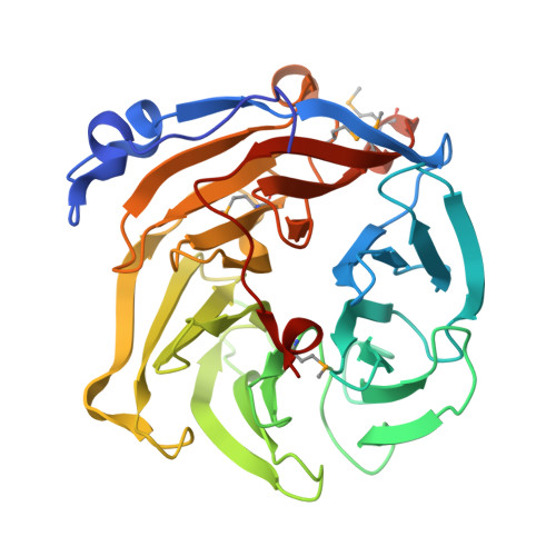
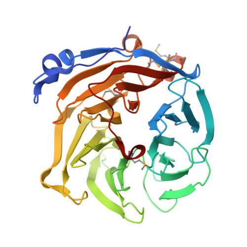
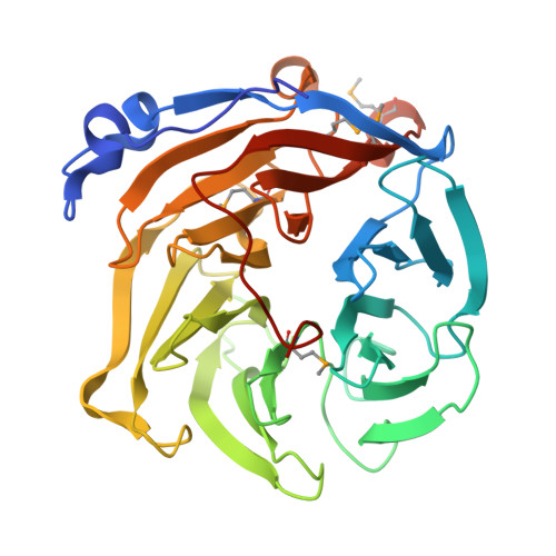
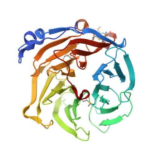
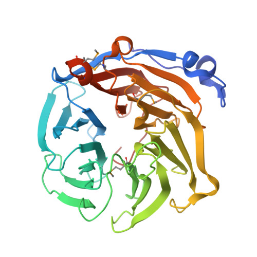
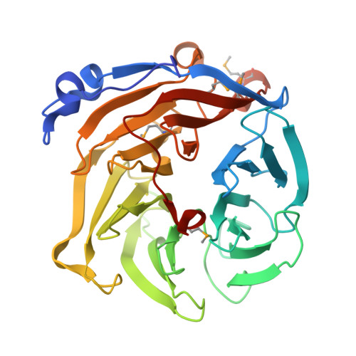
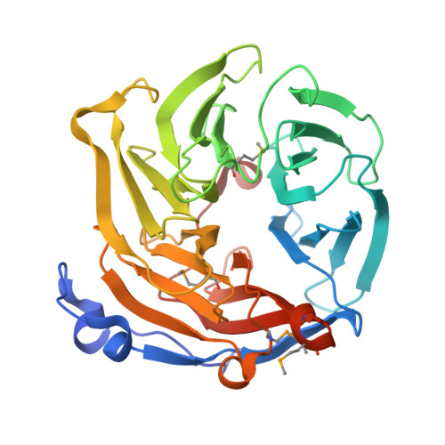
    Drp35 is a protein induced by cell wall-affecting antibiotics or detergents; it possesses calcium-dependent lactonase activity. To determine the molecular basis of the lactonase activity, we first solved the crystal structures of Drp35 with and without Ca(2+); these showed that the molecule has a six-bladed beta-propeller structure with two calcium ions bound at the center of the beta-propeller and surface region. Mutational analyses of evolutionarily conserved residues revealed that the central calcium-binding site is essential for the enzymatic activity of Drp35. Substitution of some other amino acid residues for the calcium-binding residues demonstrated the critical contributions of Glu(48), Asp(138), and Asp(236) to the enzymatic activity. Differential scanning calorimetric analysis revealed that the loss of activity of E48Q and D236N, but not D138N, was attributed to their inability to hold the calcium ion. Further structural analysis of the D138N mutant indicates that it lacks a water molecule bound to the calcium ion rather than the calcium ion itself. Based on these observations and structural information, a possible catalytic mechanism in which the calcium ion and its binding residues play direct roles was proposed for the lactonase activity of Drp35.


  • Organizational Affiliation

    Faculty of Advanced Life Sciences, Hokkaido University, Sapporo 060-0810, Japan.


Macromolecules
Find similar proteins by:  (by identity cutoff)  |  3D Structure
Entity ID: 1
MoleculeChains Sequence LengthOrganismDetailsImage
DrP35
A, B, C, D, E
A, B, C, D, E, F, G, H, I, J, K, L
333Staphylococcus aureusMutation(s): 6 
EC: 3.1.1.25 (PDB Primary Data), 3.1.1 (UniProt)
UniProt
Find proteins for Q99QV3 (Staphylococcus aureus (strain Mu50 / ATCC 700699))
Explore Q99QV3 
Go to UniProtKB:  Q99QV3
Entity Groups  
Sequence Clusters30% Identity50% Identity70% Identity90% Identity95% Identity100% Identity
UniProt GroupQ99QV3
Sequence Annotations
Expand
  • Reference Sequence
Small Molecules
Modified Residues  1 Unique
IDChains TypeFormula2D DiagramParent
MSE
Query on MSE
A, B, C, D, E
A, B, C, D, E, F, G, H, I, J, K, L
L-PEPTIDE LINKINGC5 H11 N O2 SeMET
Experimental Data & Validation

Experimental Data

  • Method: X-RAY DIFFRACTION
  • Resolution: 2.40 Å
  • R-Value Free: 0.222 
  • R-Value Work: 0.200 
  • R-Value Observed: 0.200 
  • Space Group: P 1 21 1
Unit Cell:
Length ( Å )Angle ( ˚ )
a = 86.089α = 90
b = 146.062β = 93.39
c = 151.947γ = 90
Software Package:
Software NamePurpose
CNSrefinement
HKL-2000data reduction
SCALEPACKdata scaling
SOLVEphasing

Structure Validation

View Full Validation Report


Entry History 

Deposition Data

Revision History  (Full details and data files)

  • Version 1.0: 2006-12-12
    Type: Initial release
  • Version 1.1: 2008-04-30
    Changes: Version format compliance
  • Version 1.2: 2011-07-13
    Changes: Derived calculations, Version format compliance
  • Version 1.3: 2024-10-23
    Changes: Data collection, Database references, Derived calculations, Structure summary