2DFP | pdb_00002dfp

X-RAY STRUCTURE OF AGED DI-ISOPROPYL-PHOSPHORO-FLUORIDATE (DFP) BOUND TO ACETYLCHOLINESTERASE


Experimental Data Snapshot

  • Method: X-RAY DIFFRACTION
  • Resolution: 2.30 Å
  • R-Value Free: 
    0.228 (Depositor), 0.230 (DCC) 
  • R-Value Work: 
    0.186 (Depositor), 0.190 (DCC) 

Starting Model: experimental
View more details

wwPDB Validation 3D Report Full Report

Validation slider image for 2DFP

Ligand Structure Quality Assessment 


This is version 2.2 of the entry. See complete history

Literature

Crystal structures of aged phosphonylated acetylcholinesterase: nerve agent reaction products at the atomic level.

Millard, C.B.Kryger, G.Ordentlich, A.Greenblatt, H.M.Harel, M.Raves, M.L.Segall, Y.Barak, D.Shafferman, A.Silman, I.Sussman, J.L.

(1999) Biochemistry 38: 7032-7039

  • DOI: https://doi.org/10.1021/bi982678l
  • Primary Citation Related Structures: 
    1CFJ, 1SOM, 2DFP

  • PubMed Abstract: 

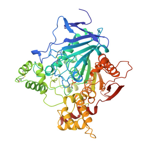
    Organophosphorus acid anhydride (OP) nerve agents are potent inhibitors which rapidly phosphonylate acetylcholinesterase (AChE) and then may undergo an internal dealkylation reaction (called "aging") to produce an OP-enzyme conjugate that cannot be reactivated. To understand the basis for irreversible inhibition, we solved the structures of aged conjugates obtained by reaction of Torpedo californica AChE (TcAChE) with diisopropylphosphorofluoridate (DFP), O-isopropylmethylphosponofluoridate (sarin), or O-pinacolylmethylphosphonofluoridate (soman) by X-ray crystallography to 2.3, 2.6, or 2.2 A resolution, respectively. The highest positive difference density peak corresponded to the OP phosphorus and was located within covalent bonding distance of the active-site serine (S200) in each structure. The OP-oxygen atoms were within hydrogen-bonding distance of four potential donors from catalytic subsites of the enzyme, suggesting that electrostatic forces significantly stabilize the aged enzyme. The active sites of aged sarin- and soman-TcAChE were essentially identical and provided structural models for the negatively charged, tetrahedral intermediate that occurs during deacylation with the natural substrate, acetylcholine. Phosphorylation with DFP caused an unexpected movement in the main chain of a loop that includes residues F288 and F290 of the TcAChE acyl pocket. This is the first major conformational change reported in the active site of any AChE-ligand complex, and it offers a structural explanation for the substrate selectivity of AChE.


  • Organizational Affiliation
    • Department of Neurobiology, Weizmann Institute of Science, Rehovot, Israel. 1imill@sgjs6.weizmann.ac.il

Macromolecule Content 

  • Total Structure Weight: 61.83 kDa 
  • Atom Count: 4,728 
  • Modeled Residue Count: 534 
  • Deposited Residue Count: 534 
  • Unique protein chains: 1

Macromolecules

Find similar proteins by:|  3D Structure
Entity ID: 1
MoleculeChains  Sequence LengthOrganismDetailsImage
PROTEIN (ACETYLCHOLINESTERASE)534Tetronarce californicaMutation(s): 0 
EC: 3.1.1.7
UniProt
Find proteins for P04058 (Tetronarce californica)
Explore P04058 
Go to UniProtKB:  P04058
Entity Groups
Sequence Clusters30% Identity50% Identity70% Identity90% Identity95% Identity100% Identity
UniProt GroupP04058
Glycosylation
Glycosylation Sites: 3
Sequence Annotations
Expand
Reference Sequence

Oligosaccharides

Help  
Entity ID: 2
MoleculeChains Length2D Diagram GlycosylationD Interactions
2-acetamido-2-deoxy-beta-D-glucopyranose-(1-4)-2-acetamido-2-deoxy-beta-D-glucopyranose
B, C
2N-Glycosylation
Glycosylation Resources
GlyTouCan: G42666HT
GlyCosmos: G42666HT
GlyGen: G42666HT

Small Molecules

Ligands 2 Unique
IDChains Name / Formula / InChI Key2D Diagram3D Interactions
NAG

Query on NAG


Download:Ideal Coordinates CCD File
D [auth A]2-acetamido-2-deoxy-beta-D-glucopyranose
C8 H15 N O6
OVRNDRQMDRJTHS-FMDGEEDCSA-N
MES

Query on MES


Download:Ideal Coordinates CCD File
E [auth A]2-(N-MORPHOLINO)-ETHANESULFONIC ACID
C6 H13 N O4 S
SXGZJKUKBWWHRA-UHFFFAOYSA-N
Modified Residues  1 Unique
IDChains TypeFormula2D DiagramParent
MIS
Query on MIS
A
L-PEPTIDE LINKINGC6 H14 N O6 PSER

Experimental Data & Validation

Experimental Data

  • Method: X-RAY DIFFRACTION
  • Resolution: 2.30 Å
  • R-Value Free:  0.228 (Depositor), 0.230 (DCC) 
  • R-Value Work:  0.186 (Depositor), 0.190 (DCC) 
Space Group: P 31 2 1
Unit Cell:
Length ( Å )Angle ( ˚ )
a = 112.636α = 90
b = 112.636β = 90
c = 136.881γ = 120
Software Package:
Software NamePurpose
DENZOdata reduction
SCALEPACKdata scaling
CNSrefinement
CNSphasing

Structure Validation

View Full Validation Report


Ligand Structure Quality Assessment 


Entry History 

Deposition Data

Revision History  (Full details and data files)

  • Version 1.0: 1999-06-28
    Type: Initial release
  • Version 1.1: 2007-10-16
    Changes: Version format compliance
  • Version 1.2: 2011-07-13
    Changes: Non-polymer description, Version format compliance
  • Version 2.0: 2020-07-29
    Type: Remediation
    Reason: Carbohydrate remediation
    Changes: Atomic model, Data collection, Derived calculations, Structure summary
  • Version 2.1: 2021-06-02
    Changes: Derived calculations, Structure summary
  • Version 2.2: 2023-08-23
    Changes: Data collection, Database references, Refinement description