2DDC | pdb_00002ddc

Unique behavior of a histidine responsible for an engineered green-to-red photoconversion process


Experimental Data Snapshot

  • Method: X-RAY DIFFRACTION
  • Resolution: 1.55 Å
  • R-Value Free: 
    0.212 (Depositor) 
  • R-Value Work: 
    0.190 (Depositor) 

Starting Model: experimental
View more details

wwPDB Validation 3D Report Full Report

Validation slider image for 2DDC

This is version 3.0 of the entry. See complete history

Literature

The E1 mechanism in photo-induced beta-elimination reactions for green-to-red conversion of fluorescent proteins.

Tsutsui, H.Shimizu, H.Mizuno, H.Nukina, N.Furuta, T.Miyawaki, A.

(2009) Chem Biol 16: 1140-1147

  • DOI: https://doi.org/10.1016/j.chembiol.2009.10.010
  • Primary Citation Related Structures: 
    2DDC, 2DDD

  • PubMed Abstract: 

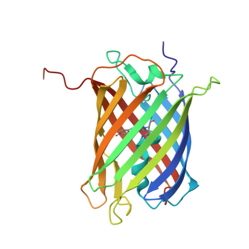
    KikGR is a fluorescent protein engineered to display green-to-red photoconvertibility that is induced by irradiation with ultraviolet or violet light. Similar to Kaede and EosFP, two naturally occurring photoconvertible proteins, KikGR contains a His(62)-Tyr(63)-Gly(64) tripeptide sequence, which forms a green chromophore that can be photoconverted to a red one via formal beta-elimination and subsequent extension of a pi-conjugated system. Using a crystallizable variant of KikGR, we determined the structures of both the green and red state at 1.55 A resolution. The double bond between His(62)-C(alpha) and His(62)-C(beta) in the red chromophore is in a cis configuration, indicating that rotation along the His(62) C(alpha)-C(beta) bond occurs following cleavage of the His(62) N(alpha)-C(alpha) bond. This structural rearrangement provides evidence that the beta-elimination reaction governing the green-to-red photoconversion of KikGR follows an E1 (elimination, unimolecular) mechanism.


  • Organizational Affiliation
    • Brain Science Institute, RIKEN, Hirosawa, Wako-city, Saitama, Japan.

Macromolecule Content 

  • Total Structure Weight: 51.51 kDa 
  • Atom Count: 4,168 
  • Modeled Residue Count: 449 
  • Deposited Residue Count: 450 
  • Unique protein chains: 1

Macromolecules

Find similar proteins by:|  3D Structure
Entity ID: 1
MoleculeChains  Sequence LengthOrganismDetailsImage
photoconvertible fluorescent protein
A, B
225Dipsastraea favusMutation(s): 6 
UniProt
Find proteins for Q53UG8 (Dipsastraea favus)
Explore Q53UG8 
Go to UniProtKB:  Q53UG8
Entity Groups
Sequence Clusters30% Identity50% Identity70% Identity90% Identity95% Identity100% Identity
UniProt GroupQ53UG8
Sequence Annotations
Expand
Reference Sequence

Small Molecules

Experimental Data & Validation

Experimental Data

  • Method: X-RAY DIFFRACTION
  • Resolution: 1.55 Å
  • R-Value Free:  0.212 (Depositor) 
  • R-Value Work:  0.190 (Depositor) 
Space Group: C 1 2 1
Unit Cell:
Length ( Å )Angle ( ˚ )
a = 97.629α = 90
b = 119.286β = 120.18
c = 49.068γ = 90
Software Package:
Software NamePurpose
HKL-2000data collection
SCALEPACKdata scaling
MOLREPphasing
CNSrefinement
HKL-2000data reduction

Structure Validation

View Full Validation Report


Entry History 

Deposition Data

Revision History  (Full details and data files)

  • Version 1.0: 2006-03-07
    Type: Initial release
  • Version 1.1: 2008-04-30
    Changes: Version format compliance
  • Version 1.2: 2011-07-13
    Changes: Version format compliance
  • Version 1.3: 2014-01-15
    Changes: Database references
  • Version 1.4: 2021-11-10
    Changes: Database references, Derived calculations
  • Version 1.5: 2023-10-25
    Changes: Data collection, Refinement description
  • Version 2.0: 2023-11-15
    Changes: Atomic model, Data collection, Derived calculations
  • Version 2.1: 2024-11-13
    Changes: Structure summary
  • Version 3.0: 2026-03-18
    Changes: Polymer sequence