2DD5 | pdb_00002dd5

Thiocyanate hydrolase (SCNase) from Thiobacillus thioparus native holo-enzyme


Experimental Data Snapshot

  • Method: X-RAY DIFFRACTION
  • Resolution: 2.00 Å
  • R-Value Free: 
    0.205 (Depositor), 0.183 (DCC) 
  • R-Value Work: 
    0.185 (Depositor), 0.184 (DCC) 
  • R-Value Observed: 
    0.185 (Depositor) 

Starting Model: experimental
View more details

wwPDB Validation 3D Report Full Report

Validation slider image for 2DD5

This is version 1.5 of the entry. See complete history

Literature

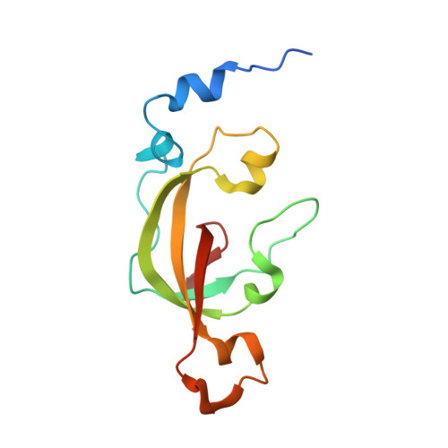
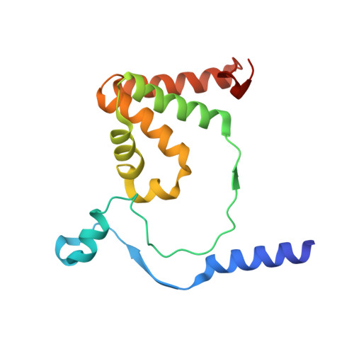
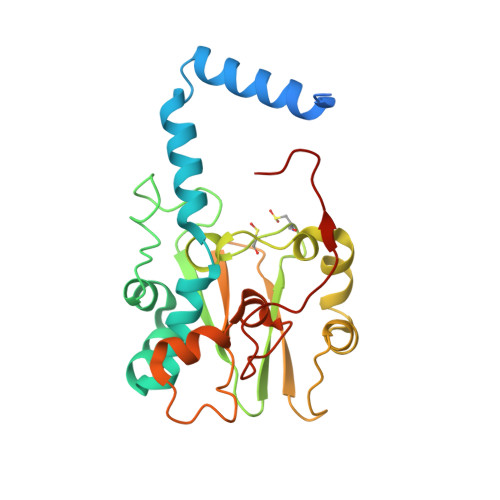
Structure of thiocyanate hydrolase: a new nitrile hydratase family protein with a novel five-coordinate cobalt(III) center.

Arakawa, T.Kawano, Y.Kataoka, S.Katayama, Y.Kamiya, N.Yohda, M.Odaka, M.

(2007) J Mol Biology 366: 1497-1509

  • DOI: https://doi.org/10.1016/j.jmb.2006.12.011
  • Primary Citation Related Structures: 
    2DD4, 2DD5

  • PubMed Abstract: 

    Thiocyanate hydrolase (SCNase) of Thiobacillus thioparus THI115 is a cobalt(III)-containing enzyme catalyzing the degradation of thiocyanate to carbonyl sulfide and ammonia. We determined the crystal structures of the apo- and native SCNases at a resolution of 2.0 A. SCNases in both forms had a conserved hetero-dodecameric structure, (alphabetagamma)(4). Four alphabetagamma hetero-trimers were structurally equivalent. One alphabetagamma hetero-trimer was composed of the core domain and the betaN domain, which was located at the center of the molecule and linked the hetero-trimers with novel quaternary interfaces. In both the apo- and native SCNases, the core domain was structurally conserved between those of iron and cobalt-types of nitrile hydratase (NHase). Native SCNase possessed the post-translationally modified cysteine ligands, gammaCys131-SO(2)H and gammaCys133-SOH like NHases. However, the low-spin cobalt(III) was found to be in the distorted square-pyramidal geometry, which had not been reported before in any protein. The size as well as the electrostatic properties of the substrate-binding pocket was totally different from NHases with respect to the charge distribution and the substrate accessibility, which rationally explains the differences in the substrate preference between SCNase and NHase.


  • Organizational Affiliation
    • Department of Biotechnology and Life Science, Graduate School of Technology, Tokyo University of Agriculture and Technology, 2-24-16 Naka-cho, Koganei, Tokyo 184-8588, Japan.

Macromolecule Content 

  • Total Structure Weight: 242.16 kDa 
  • Atom Count: 17,962 
  • Modeled Residue Count: 1,955 
  • Deposited Residue Count: 2,104 
  • Unique protein chains: 3

Macromolecules

Find similar proteins by:|  3D Structure
Entity ID: 1
MoleculeChains  Sequence LengthOrganismDetailsImage
Thiocyanate hydrolase alpha subunit
A, D, G, J
126Thiobacillus thioparusMutation(s): 0 
EC: 3.5.5.8
UniProt
Find proteins for O66187 (Thiobacillus thioparus)
Explore O66187 
Go to UniProtKB:  O66187
Entity Groups
Sequence Clusters30% Identity50% Identity70% Identity90% Identity95% Identity100% Identity
UniProt GroupO66187
Sequence Annotations
Expand
Reference Sequence
Find similar proteins by:|  3D Structure
Entity ID: 2
MoleculeChains  Sequence LengthOrganismDetailsImage
Thiocyanate hydrolase beta subunit
B, E, H, K
157Thiobacillus thioparusMutation(s): 0 
EC: 3.5.5.8
UniProt
Find proteins for O66186 (Thiobacillus thioparus)
Explore O66186 
Go to UniProtKB:  O66186
Entity Groups
Sequence Clusters30% Identity50% Identity70% Identity90% Identity95% Identity100% Identity
UniProt GroupO66186
Sequence Annotations
Expand
Reference Sequence
Find similar proteins by:|  3D Structure
Entity ID: 3
MoleculeChains  Sequence LengthOrganismDetailsImage
Thiocyanate hydrolase gamma subunit
C, F, I, L
243Thiobacillus thioparusMutation(s): 2 
EC: 3.5.5.8
UniProt
Find proteins for O66188 (Thiobacillus thioparus)
Explore O66188 
Go to UniProtKB:  O66188
Entity Groups
Sequence Clusters30% Identity50% Identity70% Identity90% Identity95% Identity100% Identity
UniProt GroupO66188
Sequence Annotations
Expand
Reference Sequence

Small Molecules

Ligands 2 Unique
IDChains Name / Formula / InChI Key2D Diagram3D Interactions
SO4

Query on SO4


Download:Ideal Coordinates CCD File
AA [auth L]
BA [auth L]
M [auth A]
O [auth C]
P [auth D]
AA [auth L],
BA [auth L],
M [auth A],
O [auth C],
P [auth D],
R [auth F],
S [auth G],
U [auth I],
V [auth I],
W [auth I],
X [auth J],
Z [auth L]
SULFATE ION
O4 S
QAOWNCQODCNURD-UHFFFAOYSA-L
3CO

Query on 3CO


Download:Ideal Coordinates CCD File
N [auth C],
Q [auth F],
T [auth I],
Y [auth L]
COBALT (III) ION
Co
JAWGVVJVYSANRY-UHFFFAOYSA-N
Modified Residues  2 Unique
IDChains TypeFormula2D DiagramParent
CSD
Query on CSD
C, F, I, L
L-PEPTIDE LINKINGC3 H7 N O4 SCYS
CSO
Query on CSO
C, F, I, L
L-PEPTIDE LINKINGC3 H7 N O3 SCYS

Experimental Data & Validation

Experimental Data

  • Method: X-RAY DIFFRACTION
  • Resolution: 2.00 Å
  • R-Value Free:  0.205 (Depositor), 0.183 (DCC) 
  • R-Value Work:  0.185 (Depositor), 0.184 (DCC) 
  • R-Value Observed: 0.185 (Depositor) 
Space Group: P 43 21 2
Unit Cell:
Length ( Å )Angle ( ˚ )
a = 170.187α = 90
b = 170.187β = 90
c = 282.469γ = 90
Software Package:
Software NamePurpose
CNSrefinement
HKL-2000data reduction
SCALEPACKdata scaling
CNSphasing

Structure Validation

View Full Validation Report


Entry History 

Deposition Data

Revision History  (Full details and data files)

  • Version 1.0: 2007-01-30
    Type: Initial release
  • Version 1.1: 2008-04-30
    Changes: Version format compliance
  • Version 1.2: 2011-07-13
    Changes: Version format compliance
  • Version 1.3: 2023-10-25
    Changes: Data collection, Database references, Derived calculations, Refinement description
  • Version 1.4: 2023-11-15
    Changes: Data collection
  • Version 1.5: 2024-10-30
    Changes: Structure summary