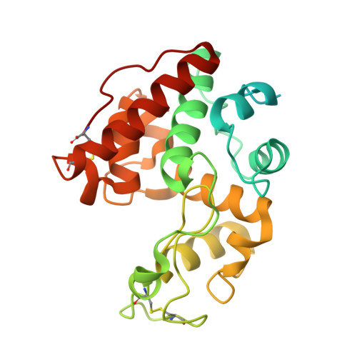
2DBT

Crystal structure of chitinase C from Streptomyces griseus HUT6037


Experimental Data Snapshot

  • Method: X-RAY DIFFRACTION
  • Resolution: 3.14 Å
  • R-Value Free: 0.225 
  • R-Value Work: 0.174 
  • R-Value Observed: 0.176 

Starting Model: experimental
View more details

wwPDB Validation   3D Report Full Report


This is version 1.4 of the entry. See complete history


Literature

Structural Studies of a Two-domain Chitinase from Streptomyces griseus HUT6037

Kezuka, Y.Ohishi, M.Itoh, Y.Watanabe, J.Mitsutomi, M.Watanabe, T.Nonaka, T.

(2006) J Mol Biol 358: 472-484

  • DOI: https://doi.org/10.1016/j.jmb.2006.02.013
  • Primary Citation of Related Structures:  
    1WVU, 1WVV, 2DBT

  • PubMed Abstract: 

    Chitinase C (ChiC) from Streptomyces griseus HUT6037 was the first glycoside hydrolase family 19 chitinase that was found in an organism other than higher plants. An N-terminal chitin-binding domain and a C-terminal catalytic domain connected by a linker peptide constitute ChiC. We determined the crystal structure of full-length ChiC, which is the only representative of the two-domain chitinases in the family. The catalytic domain has an alpha-helix-rich fold with a deep cleft containing a catalytic site, and lacks three loops on the domain surface compared with the catalytic domain of plant chitinases. The chitin-binding domain is an all-beta protein with two tryptophan residues (Trp59 and Trp60) aligned on the surface. We suggest the binding mechanism of tri-N-acetylchitotriose onto the chitin-binding domain on the basis of molecular dynamics (MD) simulations. In this mechanism, the ligand molecule binds well on the surface-exposed binding site through two stacking interactions and two hydrogen bonds and only Trp59 and Trp60 are involved in the binding. Furthermore, the flexibility of the Trp60 side-chain, which may be involved in adjusting the binding surface to fit the surface of crystalline chitin by the rotation of chi2 angle, is shown.


  • Organizational Affiliation

    Department of BioEngineering, Nagaoka University of Technology, Nagaoka, Niigata 940-2188, Japan.


Macromolecules
Find similar proteins by:  (by identity cutoff)  |  3D Structure
Entity ID: 1
MoleculeChains Sequence LengthOrganismDetailsImage
chitinase C
A, B, C
265Streptomyces griseusMutation(s): 0 
EC: 3.2.1.14
UniProt
Find proteins for O50152 (Streptomyces griseus)
Explore O50152 
Go to UniProtKB:  O50152
Entity Groups  
Sequence Clusters30% Identity50% Identity70% Identity90% Identity95% Identity100% Identity
UniProt GroupO50152
Sequence Annotations
Expand
  • Reference Sequence
Experimental Data & Validation

Experimental Data

  • Method: X-RAY DIFFRACTION
  • Resolution: 3.14 Å
  • R-Value Free: 0.225 
  • R-Value Work: 0.174 
  • R-Value Observed: 0.176 
  • Space Group: P 32 2 1
Unit Cell:
Length ( Å )Angle ( ˚ )
a = 153.116α = 90
b = 153.116β = 90
c = 90.031γ = 120
Software Package:
Software NamePurpose
REFMACrefinement
MOSFLMdata reduction
CCP4data scaling
MOLREPphasing

Structure Validation

View Full Validation Report


Entry History 

Deposition Data

Revision History  (Full details and data files)

  • Version 1.0: 2006-03-28
    Type: Initial release
  • Version 1.1: 2008-04-30
    Changes: Version format compliance
  • Version 1.2: 2011-07-13
    Changes: Version format compliance
  • Version 1.3: 2023-10-25
    Changes: Data collection, Database references, Derived calculations, Refinement description
  • Version 1.4: 2024-11-13
    Changes: Structure summary