


Structural Analysis and Molecular Interaction of Cell Division Reactivation Factor, CedA from Escherichia coli
Abe, Y., Watanabe, N., Matsuda, Y., Yoshida, Y., Katayama, T., Ueda, T.To be published.
Experimental Data Snapshot
wwPDB Validation 3D Report Full Report

Entity ID: 1 | |||||
|---|---|---|---|---|---|
| Molecule | Chains | Sequence Length | Organism | Details | Image |
| Cell division activator cedA | 62 | Escherichia coli | Mutation(s): 0 Gene Names: cedA | 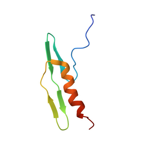 | |
UniProt | |||||
Entity Groups | |||||
| Sequence Clusters | 30% Identity50% Identity70% Identity90% Identity95% Identity100% Identity | ||||
| UniProt Group | P0AE60 | ||||
Sequence AnnotationsExpand | |||||
Reference Sequence | |||||


