2D2H | pdb_00002d2h

OpdA from Agrobacterium radiobacter with bound inhibitor trimethyl phosphate at 1.8 A resolution


Experimental Data Snapshot

  • Method: X-RAY DIFFRACTION
  • Resolution: 1.80 Å
  • R-Value Free: 
    0.214 (Depositor), 0.211 (DCC) 
  • R-Value Work: 
    0.188 (Depositor), 0.181 (DCC) 
  • R-Value Observed: 
    0.189 (Depositor) 

wwPDB Validation 3D Report Full Report

Validation slider image for 2D2H

This is version 1.4 of the entry. See complete history

Literature

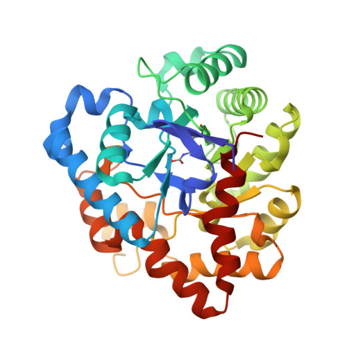
The structure of an enzyme-product complex reveals the critical role of a terminal hydroxide nucleophile in the bacterial phosphotriesterase mechanism

Jackson, C.Kim, H.K.Carr, P.D.Liu, J.W.Ollis, D.L.

(2005) Biochim Biophys Acta 1752: 56-64

  • DOI: https://doi.org/10.1016/j.bbapap.2005.06.008
  • Primary Citation Related Structures: 
    2D2G, 2D2H, 2D2J

  • PubMed Abstract: 

    A detailed understanding of the catalytic mechanism of enzymes is an important step toward improving their activity for use in biotechnology. In this paper, crystal soaking experiments and X-ray crystallography were used to analyse the mechanism of the Agrobacterium radiobacter phosphotriesterase, OpdA, an enzyme capable of detoxifying a broad range of organophosphate pesticides. The structures of OpdA complexed with ethylene glycol and the product of dimethoate hydrolysis, dimethyl thiophosphate, provide new details of the catalytic mechanism. These structures suggest that the attacking nucleophile is a terminally bound hydroxide, consistent with the catalytic mechanism of other binuclear metallophosphoesterases. In addition, a crystal structure with the potential substrate trimethyl phosphate bound non-productively demonstrates the importance of the active site cavity in orienting the substrate into an approximation of the transition state.


  • Organizational Affiliation
    • Research School of Chemistry, Australian National University, Canberra ACT 0200, Australia.

Macromolecule Content 

  • Total Structure Weight: 35.93 kDa 
  • Atom Count: 2,838 
  • Modeled Residue Count: 329 
  • Deposited Residue Count: 329 
  • Unique protein chains: 1

Macromolecules

Find similar proteins by:|  3D Structure
Entity ID: 1
MoleculeChains  Sequence LengthOrganismDetailsImage
phosphotriesterase329Agrobacterium tumefaciensMutation(s): 1 
EC: 3.1.8.1
UniProt
Find proteins for Q93LD7 (Agrobacterium tumefaciens)
Explore Q93LD7 
Go to UniProtKB:  Q93LD7
Entity Groups
Sequence Clusters30% Identity50% Identity70% Identity90% Identity95% Identity100% Identity
UniProt GroupQ93LD7
Sequence Annotations
Expand
Reference Sequence

Experimental Data & Validation

Experimental Data

  • Method: X-RAY DIFFRACTION
  • Resolution: 1.80 Å
  • R-Value Free:  0.214 (Depositor), 0.211 (DCC) 
  • R-Value Work:  0.188 (Depositor), 0.181 (DCC) 
  • R-Value Observed: 0.189 (Depositor) 
Space Group: P 31 2 1
Unit Cell:
Length ( Å )Angle ( ˚ )
a = 108.926α = 90
b = 108.926β = 90
c = 62.345γ = 120
Software Package:
Software NamePurpose
REFMACrefinement
DENZOdata reduction
SCALEPACKdata scaling
AMoREphasing

Structure Validation

View Full Validation Report


Entry History 

Deposition Data

Revision History  (Full details and data files)

  • Version 1.0: 2005-09-20
    Type: Initial release
  • Version 1.1: 2008-04-30
    Changes: Version format compliance
  • Version 1.2: 2011-07-13
    Changes: Version format compliance
  • Version 1.3: 2015-08-19
    Changes: Non-polymer description
  • Version 1.4: 2025-03-26
    Changes: Data collection, Database references, Derived calculations, Structure summary