2D1V | pdb_00002d1v

Crystal structure of DNA-binding domain of Bacillus subtilis YycF


Experimental Data Snapshot

  • Method: X-RAY DIFFRACTION
  • Resolution: 2.40 Å
  • R-Value Free: 
    0.232 (Depositor), 0.231 (DCC) 
  • R-Value Work: 
    0.228 (Depositor), 0.229 (DCC) 
  • R-Value Observed: 
    0.229 (Depositor) 

wwPDB Validation 3D Report Full Report

Validation slider image for 2D1V

This is version 1.4 of the entry. See complete history

Literature

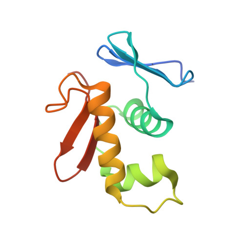
Response regulator YycF essential for bacterial growth: X-ray crystal structure of the DNA-binding domain and its PhoB-like DNA recognition motif

Okajima, T.Doi, A.Okada, A.Gotoh, Y.Tanizawa, K.Utsumi, R.

(2008) FEBS Lett 582: 3434-3438

  • DOI: https://doi.org/10.1016/j.febslet.2008.09.007
  • Primary Citation Related Structures: 
    2D1V

  • PubMed Abstract: 

    A response regulator YycF and its cognate sensor kinase YycG constitute the two-component signal transduction system essential for growth of Gram-positive bacteria with a low GC content. We have determined the X-ray crystal structure of the effector domain of Bacillus subtilis YycF involved in DNA binding. The structure, containing a winged helix-turn-helix motif, was found to be very similar to that of the response regulator PhoB from Escherichia coli. Specific binding of YycF to the PhoB-regulated alkaline phosphatase promoter was also demonstrated.


  • Organizational Affiliation
    • Institute of Scientific and Industrial Research, Osaka University, Ibaraki, Osaka 567-0047, Japan. tokajima@sanken.osaka-u.ac.jp

Macromolecule Content 

  • Total Structure Weight: 12.81 kDa 
  • Atom Count: 901 
  • Modeled Residue Count: 103 
  • Deposited Residue Count: 108 
  • Unique protein chains: 1

Macromolecules

Find similar proteins by:|  3D Structure
Entity ID: 1
MoleculeChains  Sequence LengthOrganismDetailsImage
Transcriptional regulatory protein yycF108Bacillus subtilisMutation(s): 0 
Gene Names: yycF
UniProt
Find proteins for P37478 (Bacillus subtilis (strain 168))
Explore P37478 
Go to UniProtKB:  P37478
Entity Groups
Sequence Clusters30% Identity50% Identity70% Identity90% Identity95% Identity100% Identity
UniProt GroupP37478
Sequence Annotations
Expand
Reference Sequence

Experimental Data & Validation

Experimental Data

  • Method: X-RAY DIFFRACTION
  • Resolution: 2.40 Å
  • R-Value Free:  0.232 (Depositor), 0.231 (DCC) 
  • R-Value Work:  0.228 (Depositor), 0.229 (DCC) 
  • R-Value Observed: 0.229 (Depositor) 
Space Group: C 2 2 21
Unit Cell:
Length ( Å )Angle ( ˚ )
a = 45.754α = 90
b = 65.584β = 90
c = 79.07γ = 90
Software Package:
Software NamePurpose
MOSFLMdata reduction
SCALAdata scaling
MOLREPphasing
CNSrefinement
CCP4data scaling

Structure Validation

View Full Validation Report


Entry History 

Deposition Data

Revision History  (Full details and data files)

  • Version 1.0: 2006-09-12
    Type: Initial release
  • Version 1.1: 2008-04-30
    Changes: Version format compliance
  • Version 1.2: 2011-07-13
    Changes: Version format compliance
  • Version 1.3: 2017-10-11
    Changes: Refinement description
  • Version 1.4: 2024-03-13
    Changes: Data collection, Database references