2D0P

Structure of diol dehydratase-reactivating factor in nucleotide free form


Experimental Data Snapshot

  • Method: X-RAY DIFFRACTION
  • Resolution: 3.00 Å
  • R-Value Free: 0.313 
  • R-Value Work: 0.238 
  • R-Value Observed: 0.238 

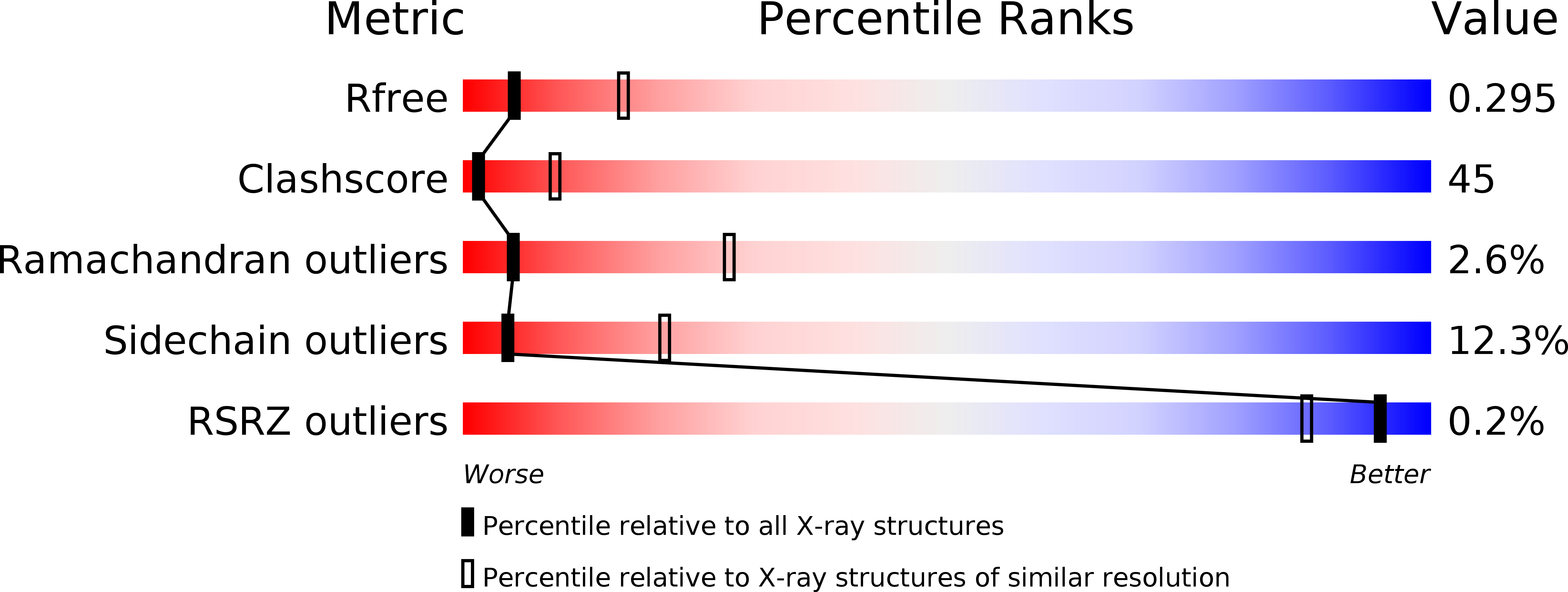
wwPDB Validation   3D Report Full Report


This is version 1.4 of the entry. See complete history


Literature

Release of a damaged cofactor from a coenzyme B12-dependent enzyme: X-ray structures of diol dehydratase-reactivating factor

Shibata, N.Mori, K.Hieda, N.Higuchi, Y.Yamanishi, M.Toraya, T.

(2005) Structure 13: 1745-1754

  • DOI: https://doi.org/10.1016/j.str.2005.08.011
  • Primary Citation of Related Structures:  
    2D0O, 2D0P

  • PubMed Abstract: 

    The crystal structures of ADP bound and nucleotide-free forms of molecular chaperone-like diol dehydratase-reactivating factor (DDR) were determined at 2.0 and 3.0 A, respectively. DDR exists as a dimer of heterodimer (alphabeta)2. The alpha subunit has four domains: ATPase domain, swiveling domain, linker domain, and insert domain. The beta subunit, composed of a single domain, has a similar fold to the beta subunit of diol dehydratase (DD). The binding of an ADP molecule to the nucleotide binding site of DDR causes a marked conformational change of the ATPase domain of the alpha subunit, which would weaken the interactions between the DDR alpha and beta subunits and make the displacement of the DDR beta subunit by DD through the beta subunit possible. The binding of the DD beta subunit to the DDR alpha subunit induces steric repulsion between the DDR alpha and DD alpha subunits that would lead to the release of a damaged cofactor from inactivated holoDD.


  • Organizational Affiliation

    Graduate School of Life Science, University of Hyogo, 3-2-1 Kouto, Ako-gun, Hyogo 678-1297, Japan.


Macromolecules
Find similar proteins by:  (by identity cutoff)  |  3D Structure
Entity ID: 1
MoleculeChains Sequence LengthOrganismDetailsImage
diol dehydratase-reactivating factor large subunit
A, C
610Klebsiella oxytocaMutation(s): 0 
UniProt
Find proteins for O68195 (Klebsiella michiganensis (strain ATCC 8724 / DSM 4798 / JCM 20051 / NBRC 3318 / NRRL B-199 / KCTC 1686 / BUCSAV 143 / CCM 1901))
Explore O68195 
Go to UniProtKB:  O68195
Entity Groups  
Sequence Clusters30% Identity50% Identity70% Identity90% Identity95% Identity100% Identity
UniProt GroupO68195
Sequence Annotations
Expand
  • Reference Sequence
Find similar proteins by:  (by identity cutoff)  |  3D Structure
Entity ID: 2
MoleculeChains Sequence LengthOrganismDetailsImage
diol dehydratase-reactivating factor small subunit
B, D
125Klebsiella oxytocaMutation(s): 0 
UniProt
Find proteins for O68196 (Klebsiella michiganensis (strain ATCC 8724 / DSM 4798 / JCM 20051 / NBRC 3318 / NRRL B-199 / KCTC 1686 / BUCSAV 143 / CCM 1901))
Explore O68196 
Go to UniProtKB:  O68196
Entity Groups  
Sequence Clusters30% Identity50% Identity70% Identity90% Identity95% Identity100% Identity
UniProt GroupO68196
Sequence Annotations
Expand
  • Reference Sequence
Experimental Data & Validation

Experimental Data

  • Method: X-RAY DIFFRACTION
  • Resolution: 3.00 Å
  • R-Value Free: 0.313 
  • R-Value Work: 0.238 
  • R-Value Observed: 0.238 
  • Space Group: P 21 21 21
Unit Cell:
Length ( Å )Angle ( ˚ )
a = 81.92α = 90
b = 85.37β = 90
c = 296.99γ = 90
Software Package:
Software NamePurpose
CNSrefinement
HKL-2000data reduction
SCALEPACKdata scaling
CNSphasing

Structure Validation

View Full Validation Report


Entry History 

Deposition Data

Revision History  (Full details and data files)

  • Version 1.0: 2006-02-28
    Type: Initial release
  • Version 1.1: 2008-04-30
    Changes: Version format compliance
  • Version 1.2: 2011-07-13
    Changes: Version format compliance
  • Version 1.3: 2020-09-09
    Changes: Derived calculations, Structure summary
  • Version 1.4: 2024-03-13
    Changes: Data collection, Database references