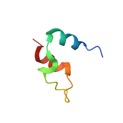
Atom-by-atom analysis of global downhill protein folding.
Sadqi, M., Fushman, D., Munoz, V.(2006) Nature 442: 317-321
- PubMed: 16799571
- DOI: https://doi.org/10.1038/nature04859
- Primary Citation of Related Structures:
2CYU - PubMed Abstract:
Protein folding is an inherently complex process involving coordination of the intricate networks of weak interactions that stabilize native three-dimensional structures. In the conventional paradigm, simple protein structures are assumed to fold in an all-or-none process that is inaccessible to experiment. Existing experimental methods therefore probe folding mechanisms indirectly. A widely used approach interprets changes in protein stability and/or folding kinetics, induced by engineered mutations, in terms of the structure of the native protein. In addition to limitations in connecting energetics with structure, mutational methods have significant experimental uncertainties and are unable to map complex networks of interactions. In contrast, analytical theory predicts small barriers to folding and the possibility of downhill folding. These theoretical predictions have been confirmed experimentally in recent years, including the observation of global downhill folding. However, a key remaining question is whether downhill folding can indeed lead to the high-resolution analysis of protein folding processes. Here we show, with the use of nuclear magnetic resonance (NMR), that the downhill protein BBL from Escherichia coli unfolds atom by atom starting from a defined three-dimensional structure. Thermal unfolding data on 158 backbone and side-chain protons out of a total of 204 provide a detailed view of the structural events during folding. This view confirms the statistical nature of folding, and exposes the interplay between hydrogen bonding, hydrophobic forces, backbone conformation and side-chain entropy. From the data we also obtain a map of the interaction network in this protein, which reveals the source of folding cooperativity. Our approach can be extended to other proteins with marginal barriers (less than 3RT), providing a new tool for the study of protein folding.
Organizational Affiliation:
Department of Chemistry and Biochemistry, and Center for Biomolecular Structure and Organization, University of Maryland, College Park, Maryland 20742, USA.


