2CW9 | pdb_00002cw9

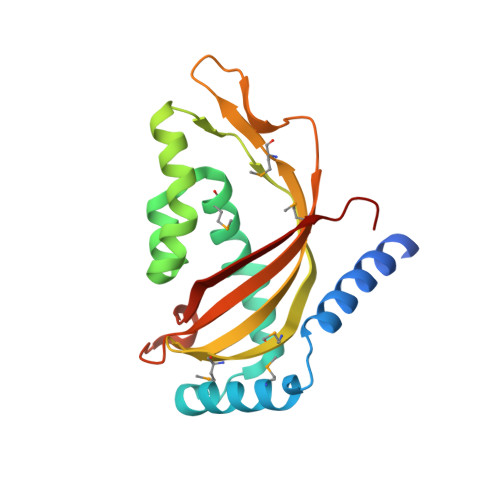
Crystal structure of human Tim44 C-terminal domain


Experimental Data Snapshot

  • Method: X-RAY DIFFRACTION
  • Resolution: 1.90 Å
  • R-Value Free: 
    0.221 (Depositor), 0.229 (DCC) 
  • R-Value Work: 
    0.218 (Depositor), 0.222 (DCC) 
  • R-Value Observed: 
    0.218 (Depositor) 

wwPDB Validation 3D Report Full Report

Validation slider image for 2CW9

This is version 1.3 of the entry. See complete history. 

Literature

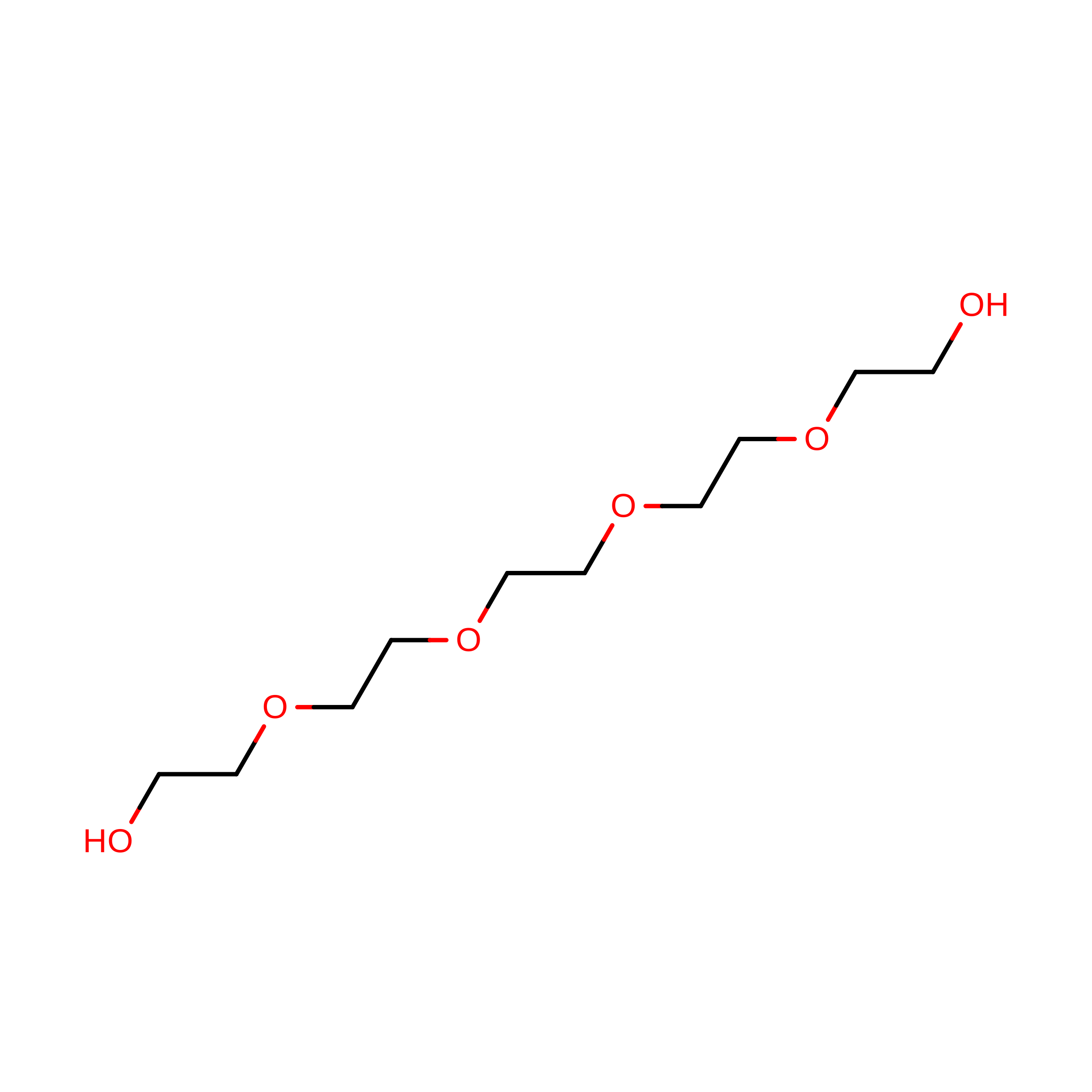
Structure of the human Tim44 C-terminal domain in complex with pentaethylene glycol: ligand-bound form.

Handa, N., Kishishita, S., Morita, S., Akasaka, R., Jin, Z., Chrzas, J., Chen, L., Liu, Z.J., Wang, B.C., Sugano, S., Tanaka, A., Terada, T., Shirouzu, M., Yokoyama, S.

(2007) Acta Crystallogr D Biol Crystallogr 63: 1225-1234

  • DOI: https://doi.org/10.1107/S0907444907051463
  • Primary Citation Related Structures: 
    2CW9

  • PubMed Abstract: 

    Familial oncocytic thyroid carcinoma is associated with a missense mutation, P308Q, in the C-terminal domain of Tim44. Tim44 is the mitochondrial inner-membrane translocase subunit and it functions as a membrane anchor for the mitochondrial heat-shock protein 70 (mtHsp70). Here, the crystal structure of the human Tim44 C-terminal domain complexed with pentaethylene glycol has been determined at 1.9 A resolution. The overall structure resembles that of the nuclear transport factor 2-like domain. In the crystal structure, pentaethylene glycol molecules are associated at two potential membrane-binding sites: the large hydrophobic cavity and the highly conserved loop between the alpha1 and alpha2 helices near Pro308. A comparison with the yeast homolog revealed that lipid binding induces conformational changes around the alpha1-alpha2 loop, leading to slippage of the alpha1 helix along the large beta-sheet. These changes may play important roles in the translocation of polypeptides across the mitochondrial inner membrane.


  • Organizational Affiliation: 
    • Protein Research Group, Genomic Sciences Center, Yokohama Institute, RIKEN, 1-7-22 Suehiro, Tsurumi, Yokohama 230-0045, Japan.

Macromolecule Content 

  • Total Structure Weight: 22.41 kDa 
  • Atom Count: 1,624 
  • Modeled Residue Count: 182 
  • Deposited Residue Count: 194 
  • Unique protein chains: 1

Macromolecules

Find similar proteins by:|  3D Structure
Entity ID: 1
MoleculeChains  Sequence LengthOrganismDetailsImage
translocase of inner mitochondrial membrane194Homo sapiensMutation(s): 0 
UniProt & NIH Common Fund Data Resources
Find proteins for O43615 (Homo sapiens)
Explore O43615 
Go to UniProtKB:  O43615
PHAROS:  O43615
GTEx:  ENSG00000104980 
Entity Groups
Sequence Clusters30% Identity50% Identity70% Identity90% Identity95% Identity100% Identity
UniProt GroupO43615
Sequence Annotations
Expand
Reference Sequence

Small Molecules

Modified Residues  1 Unique
IDChains TypeFormula2D DiagramParent
MSE
Query on MSE
A
L-PEPTIDE LINKINGC5 H11 N O2 SeMET

Experimental Data & Validation

Experimental Data

  • Method: X-RAY DIFFRACTION
  • Resolution: 1.90 Å
  • R-Value Free:  0.221 (Depositor), 0.229 (DCC) 
  • R-Value Work:  0.218 (Depositor), 0.222 (DCC) 
  • R-Value Observed: 0.218 (Depositor) 
Space Group: P 65 2 2
Unit Cell:
Length ( Å )Angle ( ˚ )
a = 108.119α = 90
b = 108.119β = 90
c = 114.056γ = 120
Software Package:
Software NamePurpose
CNSrefinement
HKL-2000data reduction
SCALEPACKdata scaling
SOLVEphasing

Structure Validation

View Full Validation Report


Entry History 

Revision History  (Full details and data files)

  • Version 1.0: 2005-12-17
    Type: Initial release
  • Version 1.1: 2007-10-30
    Changes: Version format compliance
  • Version 1.2: 2011-07-13
    Changes: Version format compliance
  • Version 1.3: 2024-11-20
    Changes: Data collection, Database references, Derived calculations, Structure summary