


Crystal structure of Phosphoglycerate Kinase from Pyrococcus horikoshii OT3
Mizutani, H., Kunishima, N.To be published.
Experimental Data Snapshot
Starting Model: experimental
View more details
Entity ID: 1 | |||||
|---|---|---|---|---|---|
| Molecule | Chains | Sequence Length | Organism | Details | Image |
| Phosphoglycerate kinase | 410 | Pyrococcus horikoshii OT3 | Mutation(s): 0 EC: 2.7.2.3 | 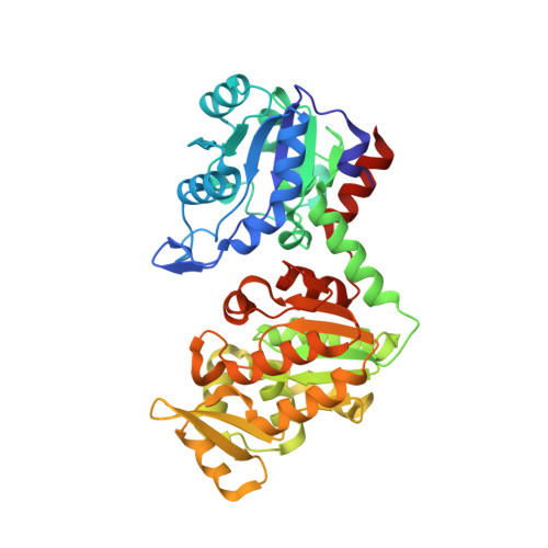 | |
UniProt | |||||
Entity Groups | |||||
| Sequence Clusters | 30% Identity50% Identity70% Identity90% Identity95% Identity100% Identity | ||||
| UniProt Group | O58965 | ||||
Sequence AnnotationsExpand | |||||
Reference Sequence | |||||
| Ligands 4 Unique | |||||
|---|---|---|---|---|---|
| ID | Chains | Name / Formula / InChI Key | 2D Diagram | 3D Interactions | |
| 3PG Download:Ideal Coordinates CCD File | D [auth A], I [auth B] | 3-PHOSPHOGLYCERIC ACID C3 H7 O7 P OSJPPGNTCRNQQC-UWTATZPHSA-N |  | ||
| MPD Download:Ideal Coordinates CCD File | F [auth A], G [auth A], K [auth B] | (4S)-2-METHYL-2,4-PENTANEDIOL C6 H14 O2 SVTBMSDMJJWYQN-YFKPBYRVSA-N |  | ||
| GOL Download:Ideal Coordinates CCD File | E [auth A], J [auth B] | GLYCEROL C3 H8 O3 PEDCQBHIVMGVHV-UHFFFAOYSA-N |  | ||
| CL Download:Ideal Coordinates CCD File | C [auth A], H [auth B] | CHLORIDE ION Cl VEXZGXHMUGYJMC-UHFFFAOYSA-M |  | ||
| Length ( Å ) | Angle ( ˚ ) |
|---|---|
| a = 49.854 | α = 90 |
| b = 123.327 | β = 95.42 |
| c = 82.943 | γ = 90 |
| Software Name | Purpose |
|---|---|
| CNS | refinement |
| HKL-2000 | data reduction |
| SCALEPACK | data scaling |
| EPMR | phasing |


