2CNN

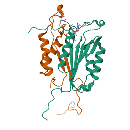
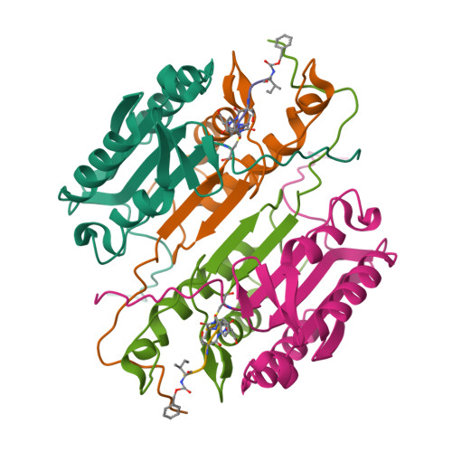
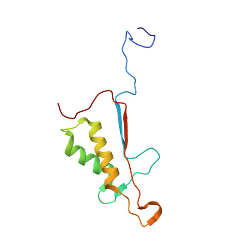
Crystal structures of caspase-3 in complex with aza-peptide epoxide inhibitors.


Experimental Data Snapshot

  • Method: X-RAY DIFFRACTION
  • Resolution: 1.70 Å
  • R-Value Free: 0.206 
  • R-Value Work: 0.183 
  • R-Value Observed: 0.183 

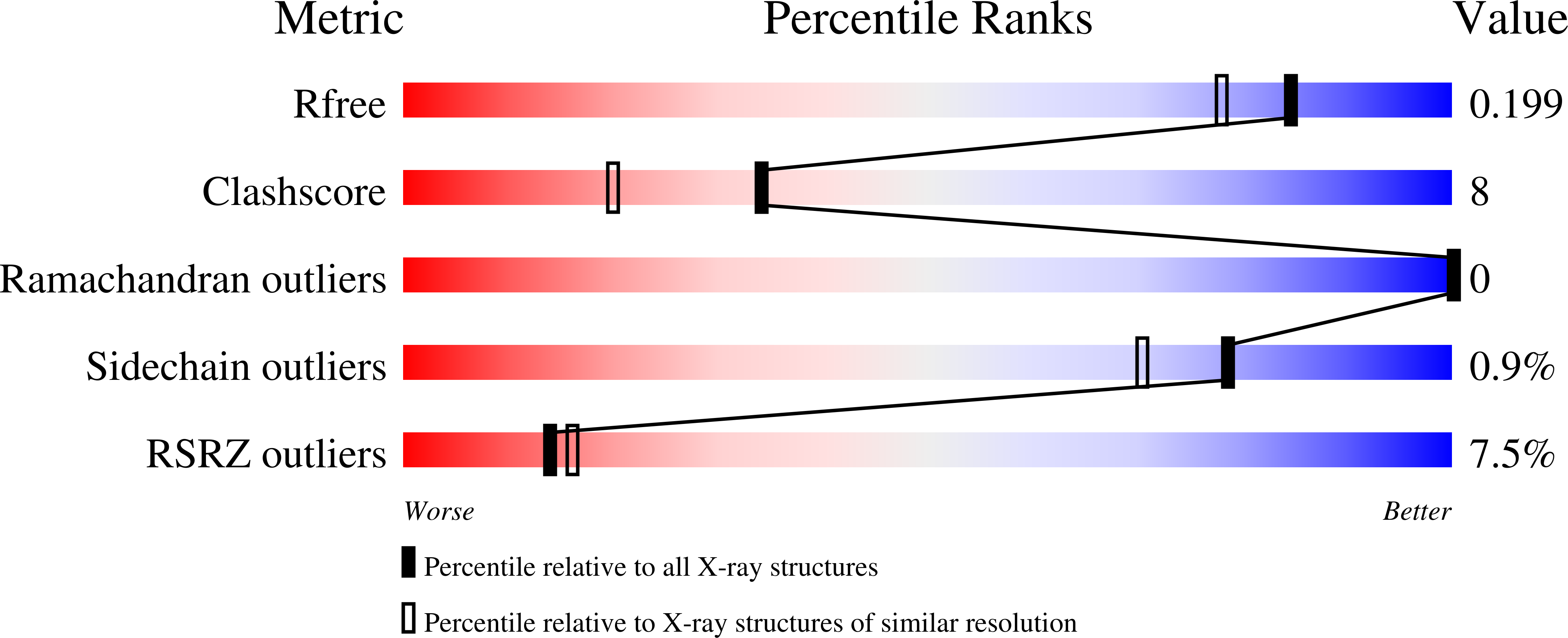
wwPDB Validation   3D Report Full Report


This is version 3.1 of the entry. See complete history


Literature

Exploring the S4 and S1 Prime Subsite Specificities in Caspase-3 with Aza-Peptide Epoxide Inhibitors

Ganesan, R.Jelakovic, S.Campbell, A.J.Li, Z.Z.Asgian, J.L.Powers M, J.C.Gruetter, G.

(2006) Biochemistry 45: 9059

  • DOI: https://doi.org/10.1021/bi060364p
  • Primary Citation of Related Structures:  
    2CDR, 2CNK, 2CNL, 2CNN, 2CNO

  • PubMed Abstract: 

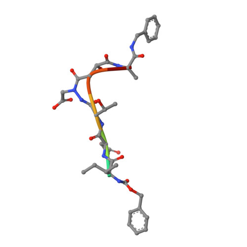
    Caspase-3 is a prototypic executioner caspase that plays a central role in apoptosis. Aza-peptide epoxides are a novel class of irreversible inhibitors that are highly specific for clan CD cysteine proteases. The five crystal structures of caspase-3-aza-peptide epoxide inhibitor complexes reported here reveal the structural basis for the mechanism of inhibition and the specificities at the S1' and the S4 subsites. Unlike the clan CA cysteine proteases, the catalytic histidine in caspase-3 plays a critical role during protonation and subsequent ring opening of the epoxide moiety and facilitates the nucleophilic attack by the active site cysteine. The nucleophilic attack takes place on the C3 carbon atom of the epoxide and results in an irreversible alkylation of the active site cysteine residue. A favorable network of hydrogen bonds involving the oxyanion hole, catalytic histidine, and the atoms in the prime site of the inhibitor enhance the binding affinity and specificity of the aza-peptide epoxide inhibitors toward caspase-3. The studies also reveal that subtle movements of the N-terminal loop of the beta-subunit occur when the P4 Asp is replaced by a P4 Ile, whereas the N-terminal loop and the safety catch Asp179 are completely disordered when the P4 Asp is replaced by P4 Cbz group.


  • Organizational Affiliation

    Department of Biochemistry, University of Zurich, Winterthurerstrasse 190, CH-8057 Zurich, Switzerland.


Macromolecules
Find similar proteins by:  (by identity cutoff)  |  3D Structure
Entity ID: 1
MoleculeChains Sequence LengthOrganismDetailsImage
Caspase-3147Homo sapiensMutation(s): 0 
Gene Names: CASP3
EC: 3.4.22.56
UniProt & NIH Common Fund Data Resources
Find proteins for P42574 (Homo sapiens)
Explore P42574 
Go to UniProtKB:  P42574
PHAROS:  P42574
GTEx:  ENSG00000164305 
Entity Groups  
Sequence Clusters30% Identity50% Identity70% Identity90% Identity95% Identity100% Identity
UniProt GroupP42574
Sequence Annotations
Expand
  • Reference Sequence
Find similar proteins by:  (by identity cutoff)  |  3D Structure
Entity ID: 2
MoleculeChains Sequence LengthOrganismDetailsImage
Caspase-3103Homo sapiensMutation(s): 0 
Gene Names: CASP3
EC: 3.4.22.56
UniProt & NIH Common Fund Data Resources
Find proteins for P42574 (Homo sapiens)
Explore P42574 
Go to UniProtKB:  P42574
PHAROS:  P42574
GTEx:  ENSG00000164305 
Entity Groups  
Sequence Clusters30% Identity50% Identity70% Identity90% Identity95% Identity100% Identity
UniProt GroupP42574
Sequence Annotations
Expand
  • Reference Sequence

Find similar proteins by:  Sequence   |   3D Structure  

Entity ID: 3
MoleculeChains Sequence LengthOrganismDetailsImage
AZA-PEPTIDE EXPOXIDE7synthetic constructMutation(s): 0 
Sequence Annotations
Expand
  • Reference Sequence
Biologically Interesting Molecules (External Reference) 1 Unique
Experimental Data & Validation

Experimental Data

  • Method: X-RAY DIFFRACTION
  • Resolution: 1.70 Å
  • R-Value Free: 0.206 
  • R-Value Work: 0.183 
  • R-Value Observed: 0.183 
  • Space Group: I 2 2 2
Unit Cell:
Length ( Å )Angle ( ˚ )
a = 67.913α = 90
b = 83.743β = 90
c = 96.211γ = 90
Software Package:
Software NamePurpose
CNSrefinement
MOSFLMdata reduction
SCALAdata scaling

Structure Validation

View Full Validation Report


Entry History 

Deposition Data

Revision History  (Full details and data files)

  • Version 1.0: 2007-05-22
    Type: Initial release
  • Version 1.1: 2011-07-13
    Changes: Atomic model, Database references, Derived calculations, Non-polymer description, Structure summary, Version format compliance
  • Version 1.2: 2012-11-30
    Changes: Other
  • Version 1.3: 2017-02-08
    Changes: Source and taxonomy
  • Version 1.4: 2017-06-28
    Changes: Data collection
  • Version 2.0: 2023-11-15
    Changes: Atomic model, Data collection, Database references, Derived calculations, Other
  • Version 3.0: 2024-01-17
    Changes: Advisory, Atomic model, Data collection, Database references, Derived calculations, Non-polymer description, Polymer sequence, Source and taxonomy, Structure summary
  • Version 3.1: 2024-11-06
    Changes: Structure summary