2CLR | pdb_00002clr

THREE DIMENSIONAL STRUCTURE OF A PEPTIDE EXTENDING OUT ONE END OF A CLASS I MHC BINDING SITE


Experimental Data Snapshot

  • Method: X-RAY DIFFRACTION
  • Resolution: 2.00 Å
  • R-Value Work: 
    0.236 (Depositor) 
  • R-Value Observed: 
    0.236 (Depositor) 

wwPDB Validation 3D Report Full Report

Validation slider image for 2CLR

This is version 1.4 of the entry. See complete history

Literature

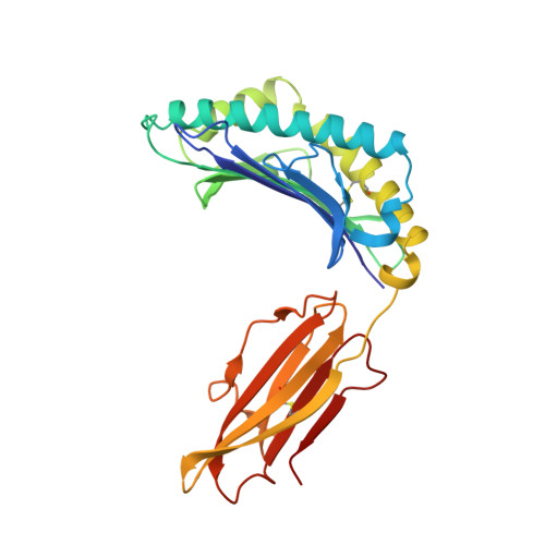
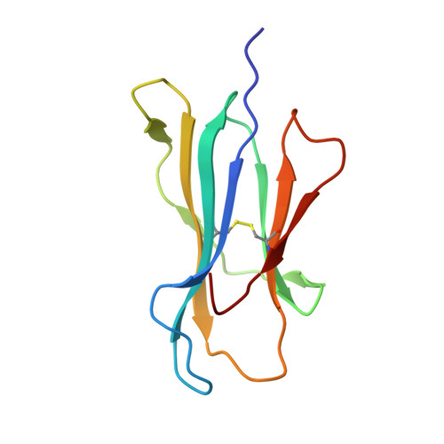
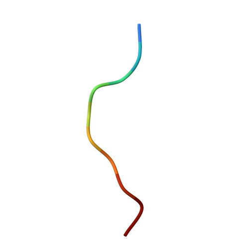
Three-dimensional structure of a peptide extending from one end of a class I MHC binding site.

Collins, E.J.Garboczi, D.N.Wiley, D.C.

(1994) Nature 371: 626-629

  • DOI: https://doi.org/10.1038/371626a0
  • Primary Citation Related Structures: 
    2CLR

  • PubMed Abstract: 

    Class I major histocompatibility complex (MHC) molecules present peptides to CD8+ T cells for immunological surveillance (reviewed in ref. 1). The structures of complexes of class I MHC molecules with octamer, nonamer and decamer peptides determined until now show a common binding mode, with both peptide termini bound in conserved pockets at the ends of the peptide binding site. Length variations were accommodated by the peptide bulging or zig-zagging in the middle. Here we describe the structure of a decamer peptide which binds with the carboxy-terminal residue positioned outside the peptide binding site. Several protein side chains have rearranged to allow the peptide to exit. The structure suggests that even longer peptides could bind. The energetic effect of the altered mode of binding has been assessed by measuring the stability of the complex to thermal denaturation. Peptides bound in this novel manner are stable at physiological temperature, raising questions about their role in T-cell recognition and their production by proteolytic processing.


  • Organizational Affiliation
    • Department of Molecular and Cellular Biology, Harvard University, Cambridge, Massachusetts 02138.

Macromolecule Content 

  • Total Structure Weight: 89.58 kDa 
  • Atom Count: 6,308 
  • Modeled Residue Count: 770 
  • Deposited Residue Count: 770 
  • Unique protein chains: 3

Macromolecules

Find similar proteins by:|  3D Structure
Entity ID: 1
MoleculeChains  Sequence LengthOrganismDetailsImage
CLASS I HISTOCOMPATIBILITY ANTIGEN (HLA-A 0201) (ALPHA CHAIN)
A, D
275Homo sapiensMutation(s): 0 
Gene Names: BETA-2-MICROGLOBULIN
UniProt & NIH Common Fund Data Resources
Find proteins for P04439 (Homo sapiens)
Explore P04439 
Go to UniProtKB:  P04439
PHAROS:  P04439
GTEx:  ENSG00000206503 
Entity Groups
Sequence Clusters30% Identity50% Identity70% Identity90% Identity95% Identity100% Identity
UniProt GroupP04439
Sequence Annotations
Expand
Reference Sequence
Find similar proteins by:|  3D Structure
Entity ID: 2
MoleculeChains  Sequence LengthOrganismDetailsImage
BETA 2-MICROGLOBULIN
B, E
100Homo sapiensMutation(s): 0 
Gene Names: BETA-2-MICROGLOBULIN
UniProt & NIH Common Fund Data Resources
Find proteins for P61769 (Homo sapiens)
Explore P61769 
Go to UniProtKB:  P61769
PHAROS:  P61769
GTEx:  ENSG00000166710 
Entity Groups
Sequence Clusters30% Identity50% Identity70% Identity90% Identity95% Identity100% Identity
UniProt GroupP61769
Sequence Annotations
Expand
Reference Sequence
Find similar proteins by:  Sequence   |   3D Structure  
Entity ID: 3
MoleculeChains  Sequence LengthOrganismDetailsImage
DECAMERIC PEPTIDE FROM CALRETICULIN
C, F
10Homo sapiensMutation(s): 0 
UniProt & NIH Common Fund Data Resources
Find proteins for P27797 (Homo sapiens)
Explore P27797 
Go to UniProtKB:  P27797
PHAROS:  P27797
GTEx:  ENSG00000179218 
Entity Groups
Sequence Clusters30% Identity50% Identity70% Identity90% Identity95% Identity100% Identity
UniProt GroupP27797
Sequence Annotations
Expand
Reference Sequence

Experimental Data & Validation

Experimental Data

  • Method: X-RAY DIFFRACTION
  • Resolution: 2.00 Å
  • R-Value Work:  0.236 (Depositor) 
  • R-Value Observed: 0.236 (Depositor) 
Space Group: P 1
Unit Cell:
Length ( Å )Angle ( ˚ )
a = 50.41α = 81.99
b = 62.909β = 76.43
c = 74.81γ = 78.06
Software Package:
Software NamePurpose
X-PLORmodel building
X-PLORrefinement
X-PLORphasing

Structure Validation

View Full Validation Report


Entry History 

Deposition Data

Revision History  (Full details and data files)

  • Version 1.0: 1995-03-31
    Type: Initial release
  • Version 1.1: 2008-03-24
    Changes: Version format compliance
  • Version 1.2: 2011-07-13
    Changes: Version format compliance
  • Version 1.3: 2024-06-05
    Changes: Data collection, Database references, Other
  • Version 1.4: 2024-10-16
    Changes: Structure summary