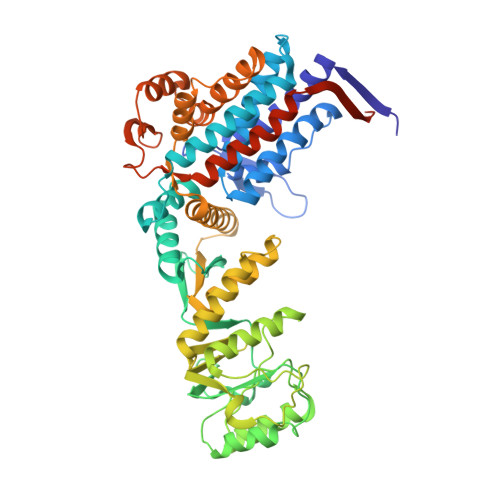
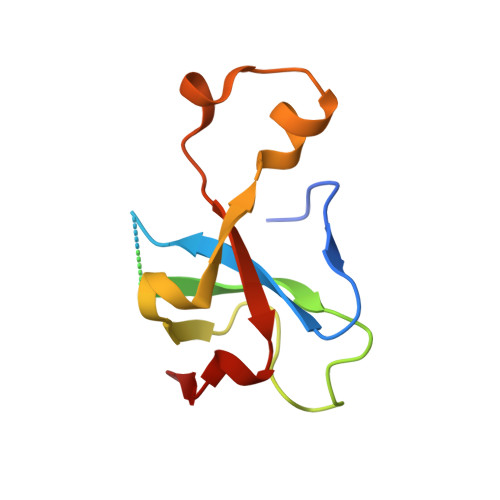
An Expanded Protein Folding Cage in the Groel-Gp31 Complex.
Clare, D.K., Bakkes, P.J., Van Heerikhuizen, H., Van Der Vies, S.M., Saibil, H.R.(2006) J Mol Biol 358: 905
- PubMed: 16549073
- DOI: https://doi.org/10.1016/j.jmb.2006.02.033
- Primary Citation of Related Structures:
2CGT - PubMed Abstract:
Bacteriophage T4 produces a GroES analogue, gp31, which cooperates with the Escherichia coli GroEL to fold its major coat protein gp23. We have used cryo-electron microscopy and image processing to obtain three-dimensional structures of the E.coli chaperonin GroEL complexed with gp31, in the presence of both ATP and ADP. The GroEL-gp31-ADP map has a resolution of 8.2 A, which allows accurate fitting of the GroEL and gp31 crystal structures. Comparison of this fitted structure with that of the GroEL-GroES-ADP structure previously determined by cryo-electron microscopy shows that the folding cage is expanded. The enlarged volume for folding is consistent with the size of the bacteriophage coat protein gp23, which is the major substrate of GroEL-gp31 chaperonin complex. At 56 kDa, gp23 is close to the maximum size limit of a polypeptide that is thought to fit inside the GroEL-GroES folding cage.
Organizational Affiliation:
School of Crystallography, Birkbeck College, University of London, Malet Street, London WC1E 7HX, UK.


