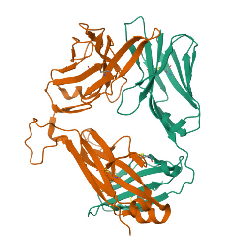
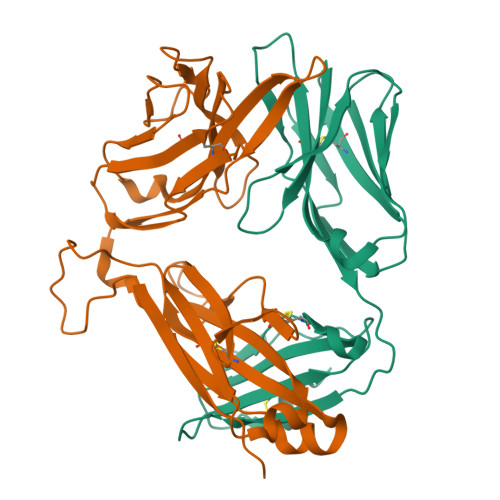
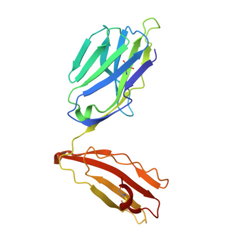
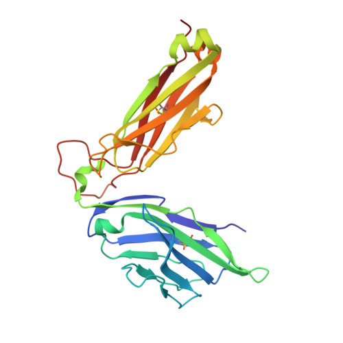
2CDF

Structure and binding kinetics of three different human CD1d-alpha- Galactosylceramide-specific T cell receptors (TCR 5E)


Experimental Data Snapshot

  • Method: X-RAY DIFFRACTION
  • Resolution: 2.25 Å
  • R-Value Free: 0.269 
  • R-Value Work: 0.188 
  • R-Value Observed: 0.192 

Starting Model: experimental
View more details

wwPDB Validation   3D Report Full Report


This is version 1.3 of the entry. See complete history


Literature

Structrue and Binding Kinetics of Three Different Human Cd1D-Alpha-Galactosylceramide-Specific T Cell Receptors

Gadola, S.D.Koch, M.Marles-Wright, J.Lissin, N.M.Sheperd, D.Matulis, G.Harlos, K.Villiger, P.M.Stuart, D.I.Jakobsen, B.K.Cerundolo, V.Jones, E.Y.

(2006) J Exp Med 203: 699

  • DOI: https://doi.org/10.1084/jem.20052369
  • Primary Citation of Related Structures:  
    2CDE, 2CDF, 2CDG

  • PubMed Abstract: 

    Invariant human TCR Valpha24-Jalpha18+/Vbeta11+ NKT cells (iNKT) are restricted by CD1d-alpha-glycosylceramides. We analyzed crystal structures and binding characteristics for an iNKT TCR plus two CD1d-alpha-GalCer-specific Vbeta11+ TCRs that use different TCR Valpha chains. The results were similar to those previously reported for MHC-peptide-specific TCRs, illustrating the versatility of the TCR platform. Docking TCR and CD1d-alpha-GalCer structures provided plausible insights into their interaction. The model supports a diagonal orientation of TCR on CD1d and suggests that complementarity determining region (CDR)3alpha, CDR3beta, and CDR1beta interact with ligands presented by CD1d, whereas CDR2beta binds to the CD1d alpha1 helix. This docking provides an explanation for the dominant usage of Vbeta11 and Vbeta8.2 chains by human and mouse iNKT cells, respectively, for recognition of CD1d-alpha-GalCer.


  • Organizational Affiliation

    Department of Rheumathology and Clinical Immunology, University of Bern, Inselspital, Berne CH-3010, Switzerland. stephan.gadola@insel.ch


Macromolecules
Find similar proteins by:  (by identity cutoff)  |  3D Structure
Entity ID: 1
MoleculeChains Sequence LengthOrganismDetailsImage
TCR 5E191Homo sapiensMutation(s): 0 
UniProt & NIH Common Fund Data Resources
Find proteins for P01848 (Homo sapiens)
Explore P01848 
Go to UniProtKB:  P01848
PHAROS:  P01848
Entity Groups  
Sequence Clusters30% Identity50% Identity70% Identity90% Identity95% Identity100% Identity
UniProt GroupP01848
Sequence Annotations
Expand
  • Reference Sequence
Find similar proteins by:  (by identity cutoff)  |  3D Structure
Entity ID: 2
MoleculeChains Sequence LengthOrganismDetailsImage
TCR 5E244Homo sapiensMutation(s): 0 
UniProt
Find proteins for P01850 (Homo sapiens)
Explore P01850 
Go to UniProtKB:  P01850
Entity Groups  
Sequence Clusters30% Identity50% Identity70% Identity90% Identity95% Identity100% Identity
UniProt GroupP01850
Sequence Annotations
Expand
  • Reference Sequence
Experimental Data & Validation

Experimental Data

  • Method: X-RAY DIFFRACTION
  • Resolution: 2.25 Å
  • R-Value Free: 0.269 
  • R-Value Work: 0.188 
  • R-Value Observed: 0.192 
  • Space Group: P 32 2 1
Unit Cell:
Length ( Å )Angle ( ˚ )
a = 64.451α = 90
b = 64.451β = 90
c = 184.977γ = 120
Software Package:
Software NamePurpose
REFMACrefinement
DENZOdata reduction
SCALEPACKdata scaling
AMoREphasing

Structure Validation

View Full Validation Report


Entry History 

Revision History  (Full details and data files)

  • Version 1.0: 2006-03-07
    Type: Initial release
  • Version 1.1: 2011-07-13
    Changes: Advisory, Version format compliance
  • Version 1.2: 2023-12-13
    Changes: Data collection, Database references, Other, Refinement description
  • Version 1.3: 2024-10-23
    Changes: Structure summary