2CC3 | pdb_00002cc3

Structure of Agrobacterium tumefaciens VirB8 protein


Experimental Data Snapshot

  • Method: X-RAY DIFFRACTION
  • Resolution: 2.20 Å
  • R-Value Free: 
    0.301 (Depositor), 0.298 (DCC) 
  • R-Value Work: 
    0.235 (Depositor), 0.234 (DCC) 
  • R-Value Observed: 
    0.239 (Depositor) 

Starting Model: experimental
View more details

wwPDB Validation 3D Report Full Report

Validation slider image for 2CC3

This is version 1.3 of the entry. See complete history

Literature

Agrobacterium Tumefaciens Virb8 Structure Reveals Potential Protein-Protein Interactions Sites.

Bailey, S.Ward, D.Middleton, R.Grossmann, J.G.Zambryski, P.C.

(2006) Proc Natl Acad Sci U S A 103: 2582

  • DOI: https://doi.org/10.1073/pnas.0511216103
  • Primary Citation Related Structures: 
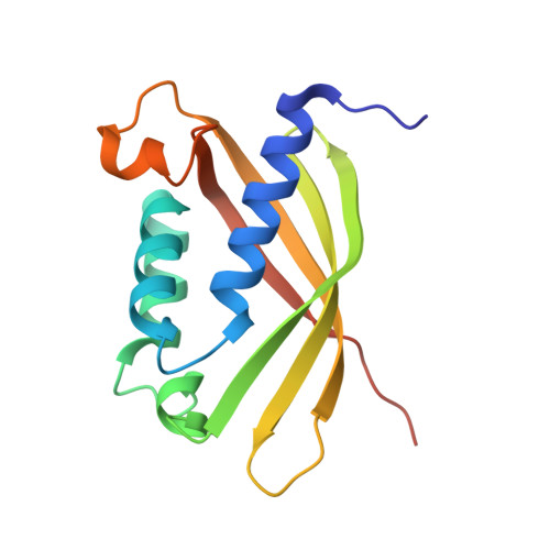
    2CC3

  • PubMed Abstract: 

    Bacterial type IV secretion systems (T4SS) translocate DNA and/or proteins to recipient cells, thus providing a mechanism for conjugative transfer of genetic material and bacterial pathogenesis. Here we describe the first structure of a core component from the archetypal Agrobacterium tumefaciens T4SS: the 2.2-A resolution crystal structure of the VirB8 periplasmic domain (pVirB8(AT)). VirB8 forms a dimer in the crystal, and we identify residues likely important for stabilization of the dimer interface. Structural comparison of pVirB8(AT) with Brucella suis VirB8 confirms that the monomers have a similar fold. In addition, the pVirB8(AT) dimer superimposes very closely on the B. suis VirB8 dimer, supporting the proposal that dimer formation in the crystal reflects self-interactions that are biologically significant. The evolutionary conservation level for each residue was obtained from a data set of 84 VirB8 homologs and projected onto the protein structure to indicate conserved surface patches that likely contact other T4SS proteins.


  • Organizational Affiliation
    • Molecular Biophysics Group, Council for the Central Laboratory of the Research Councils Daresbury Laboratory, Warrington WA4 4AD, United Kingdom.

Macromolecule Content 

  • Total Structure Weight: 33.94 kDa 
  • Atom Count: 2,477 
  • Modeled Residue Count: 288 
  • Deposited Residue Count: 300 
  • Unique protein chains: 1

Macromolecules

Find similar proteins by:|  3D Structure
Entity ID: 1
MoleculeChains  Sequence LengthOrganismDetailsImage
PROTEIN VIRB8
A, B
150Agrobacterium tumefaciensMutation(s): 0 
UniProt
Find proteins for P17798 (Agrobacterium fabrum (strain C58 / ATCC 33970))
Explore P17798 
Go to UniProtKB:  P17798
Entity Groups
Sequence Clusters30% Identity50% Identity70% Identity90% Identity95% Identity100% Identity
UniProt GroupP17798
Sequence Annotations
Expand
Reference Sequence

Experimental Data & Validation

Experimental Data

  • Method: X-RAY DIFFRACTION
  • Resolution: 2.20 Å
  • R-Value Free:  0.301 (Depositor), 0.298 (DCC) 
  • R-Value Work:  0.235 (Depositor), 0.234 (DCC) 
  • R-Value Observed: 0.239 (Depositor) 
Space Group: C 1 2 1
Unit Cell:
Length ( Å )Angle ( ˚ )
a = 112.269α = 90
b = 72.349β = 110.66
c = 48.844γ = 90
Software Package:
Software NamePurpose
REFMACrefinement
MOSFLMdata reduction
SCALAdata scaling
MOLREPphasing

Structure Validation

View Full Validation Report


Entry History 

Deposition Data

Revision History  (Full details and data files)

  • Version 1.0: 2006-01-30
    Type: Initial release
  • Version 1.1: 2011-05-08
    Changes: Version format compliance
  • Version 1.2: 2011-07-13
    Changes: Version format compliance
  • Version 1.3: 2023-12-13
    Changes: Data collection, Database references, Other, Refinement description