2C6G | pdb_00002c6g

Membrane-bound glutamate carboxypeptidase II (GCPII) with bound glutamate


Experimental Data Snapshot

  • Method: X-RAY DIFFRACTION
  • Resolution: 2.20 Å
  • R-Value Free: 
    0.228 (Depositor), 0.230 (DCC) 
  • R-Value Work: 
    0.186 (Depositor), 0.193 (DCC) 
  • R-Value Observed: 
    0.187 (Depositor) 

wwPDB Validation 3D Report Full Report

Validation slider image for 2C6G

Ligand Structure Quality Assessment 


This is version 2.1 of the entry. See complete history

Literature

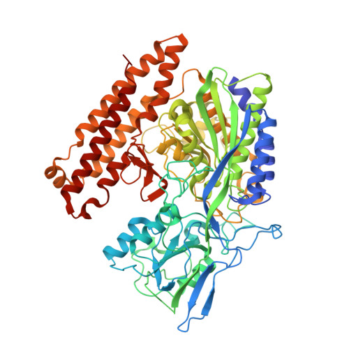
Structure of glutamate carboxypeptidase II, a drug target in neuronal damage and prostate cancer.

Mesters, J.R.Barinka, C.Li, W.Tsukamoto, T.Majer, P.Slusher, B.S.Konvalinka, J.Hilgenfeld, R.

(2006) EMBO J 25: 1375-1384

  • DOI: https://doi.org/10.1038/sj.emboj.7600969
  • Primary Citation Related Structures: 
    2C6C, 2C6G, 2C6P

  • PubMed Abstract: 

    Membrane-bound glutamate carboxypeptidase II (GCPII) is a zinc metalloenzyme that catalyzes the hydrolysis of the neurotransmitter N-acetyl-L-aspartyl-L-glutamate (NAAG) to N-acetyl-L-aspartate and L-glutamate (which is itself a neurotransmitter). Potent and selective GCPII inhibitors have been shown to decrease brain glutamate and provide neuroprotection in preclinical models of stroke, amyotrophic lateral sclerosis, and neuropathic pain. Here, we report crystal structures of the extracellular part of GCPII in complex with both potent and weak inhibitors and with glutamate, the product of the enzyme's hydrolysis reaction, at 2.0, 2.4, and 2.2 A resolution, respectively. GCPII folds into three domains: protease-like, apical, and C-terminal. All three participate in substrate binding, with two of them directly involved in C-terminal glutamate recognition. One of the carbohydrate moieties of the enzyme is essential for homodimer formation of GCPII. The three-dimensional structures presented here reveal an induced-fit substrate-binding mode of this key enzyme and provide essential information for the design of GCPII inhibitors useful in the treatment of neuronal diseases and prostate cancer.


  • Organizational Affiliation
    • Institute of Biochemistry, Center for Structural and Cell Biology in Medicine, University of Lübeck, Lübeck, Germany.

Macromolecule Content 

  • Total Structure Weight: 82.23 kDa 
  • Atom Count: 5,571 
  • Modeled Residue Count: 679 
  • Deposited Residue Count: 707 
  • Unique protein chains: 1

Macromolecules

Find similar proteins by:|  3D Structure
Entity ID: 1
MoleculeChains  Sequence LengthOrganismDetailsImage
GLUTAMATE CARBOXYPEPTIDASE II707Homo sapiensMutation(s): 0 
EC: 3.4.17.21
UniProt & NIH Common Fund Data Resources
Find proteins for Q04609 (Homo sapiens)
Explore Q04609 
Go to UniProtKB:  Q04609
PHAROS:  Q04609
GTEx:  ENSG00000086205 
Entity Groups
Sequence Clusters30% Identity50% Identity70% Identity90% Identity95% Identity100% Identity
UniProt GroupQ04609
Glycosylation
Glycosylation Sites: 6Go to GlyGen: Q04609-1
Sequence Annotations
Expand
Reference Sequence

Oligosaccharides

Help  
Entity ID: 2
MoleculeChains Length2D Diagram GlycosylationD Interactions
2-acetamido-2-deoxy-beta-D-glucopyranose-(1-4)-2-acetamido-2-deoxy-beta-D-glucopyranose
B, C
2N-Glycosylation
Glycosylation Resources
GlyTouCan: G42666HT
GlyCosmos: G42666HT
GlyGen: G42666HT
Entity ID: 3
MoleculeChains Length2D Diagram GlycosylationD Interactions
alpha-D-mannopyranose-(1-3)-beta-D-mannopyranose-(1-4)-2-acetamido-2-deoxy-beta-D-glucopyranose-(1-4)-2-acetamido-2-deoxy-beta-D-glucopyranose
D
4N-Glycosylation
Glycosylation Resources
GlyTouCan: G81315DD
GlyCosmos: G81315DD
GlyGen: G81315DD

Small Molecules

Ligands 5 Unique
IDChains Name / Formula / InChI Key2D Diagram3D Interactions
NAG

Query on NAG


Download:Ideal Coordinates CCD File
J [auth A],
K [auth A],
L [auth A]
2-acetamido-2-deoxy-beta-D-glucopyranose
C8 H15 N O6
OVRNDRQMDRJTHS-FMDGEEDCSA-N
GLU

Query on GLU


Download:Ideal Coordinates CCD File
E [auth A]GLUTAMIC ACID
C5 H9 N O4
WHUUTDBJXJRKMK-VKHMYHEASA-N
ZN

Query on ZN


Download:Ideal Coordinates CCD File
F [auth A],
G [auth A]
ZINC ION
Zn
PTFCDOFLOPIGGS-UHFFFAOYSA-N
CA

Query on CA


Download:Ideal Coordinates CCD File
H [auth A]CALCIUM ION
Ca
BHPQYMZQTOCNFJ-UHFFFAOYSA-N
CL

Query on CL


Download:Ideal Coordinates CCD File
I [auth A]CHLORIDE ION
Cl
VEXZGXHMUGYJMC-UHFFFAOYSA-M

Experimental Data & Validation

Experimental Data

  • Method: X-RAY DIFFRACTION
  • Resolution: 2.20 Å
  • R-Value Free:  0.228 (Depositor), 0.230 (DCC) 
  • R-Value Work:  0.186 (Depositor), 0.193 (DCC) 
  • R-Value Observed: 0.187 (Depositor) 
Space Group: I 2 2 2
Unit Cell:
Length ( Å )Angle ( ˚ )
a = 102.435α = 90
b = 130.428β = 90
c = 159.775γ = 90
Software Package:
Software NamePurpose
REFMACrefinement
DENZOdata reduction
SCALEPACKdata scaling

Structure Validation

View Full Validation Report


Ligand Structure Quality Assessment 


Entry History 

Deposition Data

Revision History  (Full details and data files)

  • Version 1.0: 2006-02-15
    Type: Initial release
  • Version 1.1: 2011-05-08
    Changes: Version format compliance
  • Version 1.2: 2011-07-13
    Changes: Version format compliance
  • Version 1.3: 2018-06-13
    Changes: Data collection, Database references, Refinement description, Structure summary
  • Version 1.4: 2019-07-10
    Changes: Data collection, Derived calculations
  • Version 1.5: 2019-07-24
    Changes: Data collection
  • Version 2.0: 2020-07-29
    Type: Remediation
    Reason: Carbohydrate remediation
    Changes: Advisory, Atomic model, Data collection, Derived calculations, Other, Structure summary
  • Version 2.1: 2024-11-06
    Changes: Data collection, Database references, Derived calculations, Structure summary