2C5U

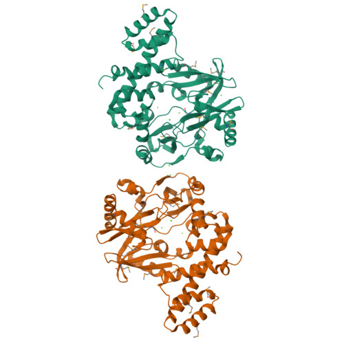
T4 RNA Ligase (Rnl1) Crystal Structure


Experimental Data Snapshot

  • Method: X-RAY DIFFRACTION
  • Resolution: 2.21 Å
  • R-Value Free: 0.258 
  • R-Value Work: 0.198 
  • R-Value Observed: 0.198 

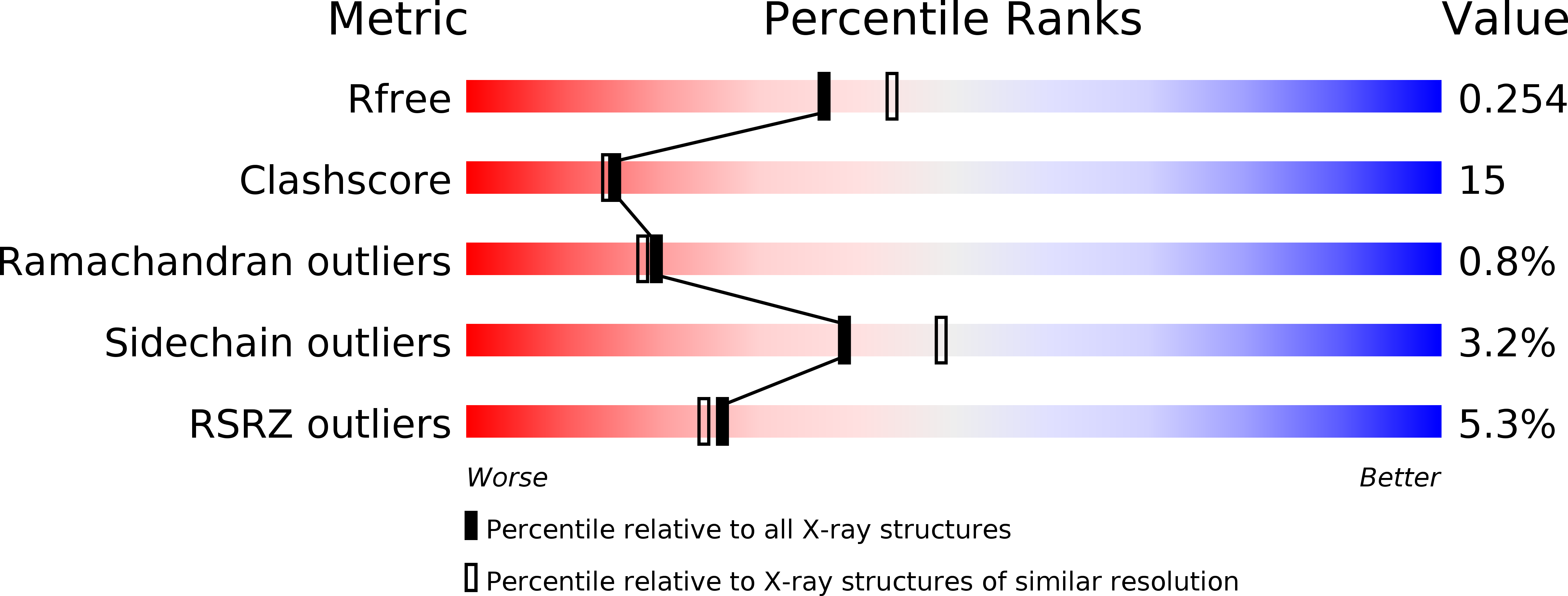
wwPDB Validation   3D Report Full Report


Ligand Structure Quality Assessment 


This is version 1.2 of the entry. See complete history


Literature

Molecular Architecture and Ligand Recognition Determinants for T4 RNA Ligase

El Omari, K.Ren, J.Bird, L.E.Bona, M.K.Klarmann, G.Legrice, S.F.J.Stammers, D.K.

(2006) J Biol Chem 281: 1573

  • DOI: https://doi.org/10.1074/jbc.M509658200
  • Primary Citation of Related Structures:  
    2C5U

  • PubMed Abstract: 

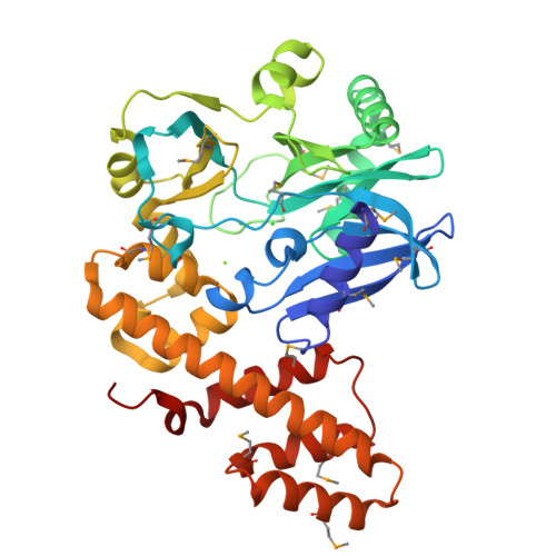
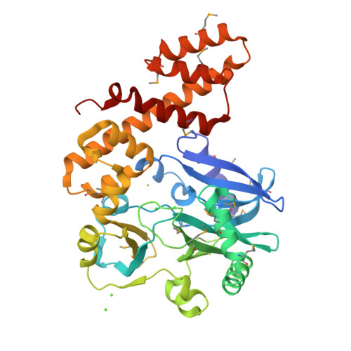
    RNA ligase type 1 from bacteriophage T4 (Rnl1) is involved in countering a host defense mechanism by repairing 5'-PO4 and 3'-OH groups in tRNA(Lys). Rnl1 is widely used as a reagent in molecular biology. Although many structures for DNA ligases are available, only fragments of RNA ligases such as Rnl2 are known. We report the first crystal structure of a complete RNA ligase, Rnl1, in complex with adenosine 5'-(alpha,beta-methylenetriphosphate) (AMPcPP). The N-terminal domain is related to the equivalent region of DNA ligases and Rnl2 and binds AMPcPP but with further interactions from the additional N-terminal 70 amino acids in Rnl1 (via Tyr37 and Arg54) and the C-terminal domain (Gly269 and Asp272). The active site contains two metal ions, consistent with the two-magnesium ion catalytic mechanism. The C-terminal domain represents a new all alpha-helical fold and has a charge distribution and architecture for helix-nucleic acid groove interaction compatible with tRNA binding.


  • Organizational Affiliation

    Division of Structural Biology, The Wellcome Trust Centre for Human Genetics, University of Oxford, Roosevelt Drive, Oxford, OX3 7BN, UK.


Macromolecules
Find similar proteins by:  (by identity cutoff)  |  3D Structure
Entity ID: 1
MoleculeChains Sequence LengthOrganismDetailsImage
RNA LIGASE
A, B
375Tequatrovirus T4Mutation(s): 0 
EC: 6.5.1.3
UniProt
Find proteins for P00971 (Enterobacteria phage T4)
Explore P00971 
Go to UniProtKB:  P00971
Entity Groups  
Sequence Clusters30% Identity50% Identity70% Identity90% Identity95% Identity100% Identity
UniProt GroupP00971
Sequence Annotations
Expand
  • Reference Sequence
Small Molecules
Ligands 4 Unique
IDChains Name / Formula / InChI Key2D Diagram3D Interactions
APC
Query on APC

Download Ideal Coordinates CCD File 
C [auth A],
H [auth B]
DIPHOSPHOMETHYLPHOSPHONIC ACID ADENOSYL ESTER
C11 H18 N5 O12 P3
CAWZRIXWFRFUQB-IOSLPCCCSA-N
CA
Query on CA

Download Ideal Coordinates CCD File 
D [auth A],
J [auth B],
K [auth B],
L [auth B]
CALCIUM ION
Ca
BHPQYMZQTOCNFJ-UHFFFAOYSA-N
CL
Query on CL

Download Ideal Coordinates CCD File 
F [auth A],
G [auth A],
M [auth B]
CHLORIDE ION
Cl
VEXZGXHMUGYJMC-UHFFFAOYSA-M
MG
Query on MG

Download Ideal Coordinates CCD File 
E [auth A],
I [auth B]
MAGNESIUM ION
Mg
JLVVSXFLKOJNIY-UHFFFAOYSA-N
Modified Residues  1 Unique
IDChains TypeFormula2D DiagramParent
MSE
Query on MSE
A, B
L-PEPTIDE LINKINGC5 H11 N O2 SeMET
Experimental Data & Validation

Experimental Data

  • Method: X-RAY DIFFRACTION
  • Resolution: 2.21 Å
  • R-Value Free: 0.258 
  • R-Value Work: 0.198 
  • R-Value Observed: 0.198 
  • Space Group: P 1 21 1
Unit Cell:
Length ( Å )Angle ( ˚ )
a = 105.05α = 90
b = 39.87β = 117.27
c = 108.54γ = 90
Software Package:
Software NamePurpose
CNSrefinement
DENZOdata reduction
SCALEPACKdata scaling
SHELXphasing

Structure Validation

View Full Validation Report


Ligand Structure Quality Assessment 


Entry History 

Deposition Data

Revision History  (Full details and data files)

  • Version 1.0: 2005-11-04
    Type: Initial release
  • Version 1.1: 2012-09-12
    Changes: Database references, Derived calculations, Non-polymer description, Other, Refinement description, Structure summary, Version format compliance
  • Version 1.2: 2024-10-23
    Changes: Data collection, Database references, Derived calculations, Other, Structure summary