2C45 | pdb_00002c45

NATIVE PRECURSOR OF PYRUVOYL DEPENDENT ASPARTATE DECARBOXYLASE


Experimental Data Snapshot

  • Method: X-RAY DIFFRACTION
  • Resolution: 2.99 Å
  • R-Value Free: 
    0.308 (Depositor), 0.292 (DCC) 
  • R-Value Work: 
    0.265 (Depositor), 0.273 (DCC) 
  • R-Value Observed: 
    0.265 (Depositor) 

Starting Model: experimental
View more details

wwPDB Validation 3D Report Full Report

Validation slider image for 2C45

This is version 1.6 of the entry. See complete history

Literature

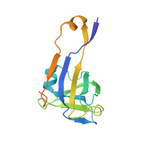
Crystal structure of uncleaved L-aspartate-alpha-decarboxylase from Mycobacterium tuberculosis.

Gopalan, G.Chopra, S.Ranganathan, A.Swaminathan, K.

(2006) Proteins 65: 796-802

  • DOI: https://doi.org/10.1002/prot.21126
  • Primary Citation Related Structures: 
    2C45

  • PubMed Abstract: 

    L-aspartate-alpha-decarboxylase (ADC) is a critical regulatory enzyme in the pantothenate biosynthetic pathway and belongs to a small class of self-cleaving and pyruvoyl-dependent amino acid decarboxylases. The expression level of ADC in Mycobacterium tuberculosis (Mtb) was confirmed by cDNA analysis, immunoblotting with an anti-ADC polyclonal antibody using whole cell lysate and immunoelectron microscopy. The recombinant ADC proenzyme from Mycobacterium tuberculosis (MtbADC) was overexpressed in E. coli and the protein structure was determined at 2.99 A resolution. The proteins fold into the double-psi beta-barrel structure. The subunits of the two tetramers (there are eight ADC molecules in the asymmetric unit) form pseudo fourfold rotational symmetry, similar to the E. coli ADC proenzyme structure. As pantothenate is synthesized in microorganisms, plants, and fungi but not in animals, structure elucidation of Mtb ADC is of substantial interest for structure-based drug development.


  • Organizational Affiliation
    • Department of Biological Sciences, National University of Singapore, Singapore 117543.

Macromolecule Content 

  • Total Structure Weight: 119.21 kDa 
  • Atom Count: 6,881 
  • Modeled Residue Count: 912 
  • Deposited Residue Count: 1,112 
  • Unique protein chains: 1

Macromolecules

Find similar proteins by:|  3D Structure
Entity ID: 1
MoleculeChains  Sequence LengthOrganismDetailsImage
Aspartate 1-decarboxylase
A, B, C, D, E
A, B, C, D, E, F, G, H
139Mycobacterium tuberculosisMutation(s): 0 
Gene Names: panDRv3601cMTCY07H7B.21
EC: 4.1.1.11
UniProt
Find proteins for P9WIL3 (Mycobacterium tuberculosis (strain ATCC 25618 / H37Rv))
Explore P9WIL3 
Go to UniProtKB:  P9WIL3
Entity Groups
Sequence Clusters30% Identity50% Identity70% Identity90% Identity95% Identity100% Identity
UniProt GroupP9WIL3
Sequence Annotations
Expand
Reference Sequence

Experimental Data & Validation

Experimental Data

  • Method: X-RAY DIFFRACTION
  • Resolution: 2.99 Å
  • R-Value Free:  0.308 (Depositor), 0.292 (DCC) 
  • R-Value Work:  0.265 (Depositor), 0.273 (DCC) 
  • R-Value Observed: 0.265 (Depositor) 
Space Group: P 31
Unit Cell:
Length ( Å )Angle ( ˚ )
a = 150.1α = 90
b = 150.1β = 90
c = 60.19γ = 120
Software Package:
Software NamePurpose
CNSrefinement
DENZOdata reduction
SCALEPACKdata scaling

Structure Validation

View Full Validation Report


Entry History 

Deposition Data

Revision History  (Full details and data files)

  • Version 1.0: 2007-03-06
    Type: Initial release
  • Version 1.1: 2011-05-07
    Changes: Version format compliance
  • Version 1.2: 2011-07-13
    Changes: Version format compliance
  • Version 1.3: 2018-01-24
    Changes: Database references
  • Version 1.4: 2018-12-12
    Changes: Data collection, Database references, Source and taxonomy, Structure summary
  • Version 1.5: 2019-05-08
    Changes: Data collection, Experimental preparation
  • Version 1.6: 2023-12-13
    Changes: Data collection, Database references, Refinement description