2C37 | pdb_00002c37

RNASE PH CORE OF THE ARCHAEAL EXOSOME IN COMPLEX WITH U8 RNA


Experimental Data Snapshot

  • Method: X-RAY DIFFRACTION
  • Resolution: 2.80 Å
  • R-Value Free: 
    0.282 (Depositor), 0.285 (DCC) 
  • R-Value Work: 
    0.266 (Depositor), 0.272 (DCC) 
  • R-Value Observed: 
    0.267 (Depositor) 

Starting Model: experimental
View more details

wwPDB Validation 3D Report Full Report

Validation slider image for 2C37

Ligand Structure Quality Assessment 


This is version 2.1 of the entry. See complete history

Literature

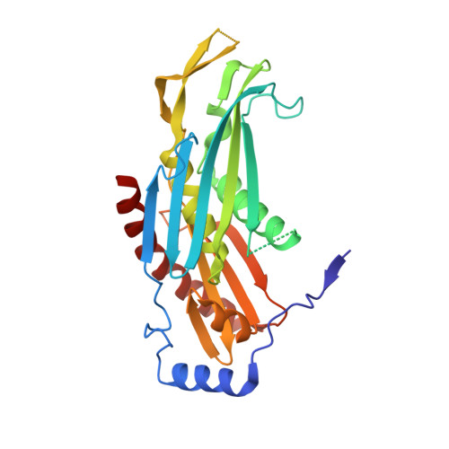
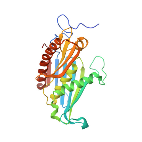
Structural Basis of 3' End RNA Recognition and Exoribonucleolytic Cleavage by an Exosome Rnase Ph Core.

Lorentzen, E.Conti, E.

(2005) Mol Cell 20: 473

  • DOI: https://doi.org/10.1016/j.molcel.2005.10.020
  • Primary Citation Related Structures: 
    2C37, 2C38, 2C39

  • PubMed Abstract: 

    The exosome is a macromolecular complex that plays fundamental roles in the biogenesis and turnover of a large number of RNA species. Here we report the crystal structures of the Rrp41-Rrp42 core complex of the S. solfataricus exosome bound to short single-stranded RNAs and to ADP. The RNA binding cleft recognizes four nucleotides in a sequence-unspecific manner, mainly by electrostatic interactions with the phosphate groups. Interactions at the 2' hydroxyls of the sugars provide specificity for RNA over DNA. The structures show both the bound substrate and the cleaved product of the reaction, suggesting a catalytic mechanism for the 3'-5' phosphorolytic activity of the exosome.


  • Organizational Affiliation
    • European Molecular Biology Laboratory, Meyerhofstrasse 1, D-69117 Heidelberg, Germany.

Macromolecule Content 

  • Total Structure Weight: 702.87 kDa 
  • Atom Count: 46,570 
  • Modeled Residue Count: 6,044 
  • Deposited Residue Count: 6,276 
  • Unique protein chains: 2

Macromolecules

Find similar proteins by:|  3D Structure
Entity ID: 1
MoleculeChains  Sequence LengthOrganismDetailsImage
PROBABLE EXOSOME COMPLEX EXONUCLEASE 2
A, C, E, G, I
A, C, E, G, I, K, M, O, Q, S, U, W
275Saccharolobus solfataricusMutation(s): 0 
EC: 3.1.13
UniProt
Find proteins for Q9UXC0 (Saccharolobus solfataricus (strain ATCC 35092 / DSM 1617 / JCM 11322 / P2))
Explore Q9UXC0 
Go to UniProtKB:  Q9UXC0
Entity Groups
Sequence Clusters30% Identity50% Identity70% Identity90% Identity95% Identity100% Identity
UniProt GroupQ9UXC0
Sequence Annotations
Expand
Reference Sequence
Find similar proteins by:|  3D Structure
Entity ID: 2
MoleculeChains  Sequence LengthOrganismDetailsImage
PROBABLE EXOSOME COMPLEX EXONUCLEASE 1
B, D, F, H, J
B, D, F, H, J, L, N, P, R, T, V, X
248Saccharolobus solfataricusMutation(s): 0 
EC: 3.1.13
UniProt
Find proteins for Q9UXC2 (Saccharolobus solfataricus (strain ATCC 35092 / DSM 1617 / JCM 11322 / P2))
Explore Q9UXC2 
Go to UniProtKB:  Q9UXC2
Entity Groups
Sequence Clusters30% Identity50% Identity70% Identity90% Identity95% Identity100% Identity
UniProt GroupQ9UXC2
Sequence Annotations
Expand
Reference Sequence

Small Molecules

Ligands 4 Unique
IDChains Name / Formula / InChI Key2D Diagram3D Interactions
U5P

Query on U5P


Download:Ideal Coordinates CCD File
AB [auth U]
BA [auth B]
BB [auth U]
CA [auth B]
CB [auth U]
AB [auth U],
BA [auth B],
BB [auth U],
CA [auth B],
CB [auth U],
EA [auth E],
EB [auth V],
FA [auth E],
GA [auth E],
GB [auth W],
HB [auth W],
IA [auth F],
IB [auth W],
JA [auth G],
KA [auth G],
KB [auth X],
MA [auth H],
NA [auth I],
OA [auth I],
QA [auth J],
SA [auth M],
TA [auth M],
VA [auth N],
Y [auth A],
Z [auth A]
URIDINE-5'-MONOPHOSPHATE
C9 H13 N2 O9 P
DJJCXFVJDGTHFX-XVFCMESISA-N
RP5

Query on RP5


Download:Ideal Coordinates CCD File
WA [auth N]5-O-phosphono-beta-D-ribofuranose
C5 H11 O8 P
KTVPXOYAKDPRHY-TXICZTDVSA-N
CL

Query on CL


Download:Ideal Coordinates CCD File
AA [auth B]
DA [auth D]
DB [auth V]
HA [auth F]
JB [auth X]
AA [auth B],
DA [auth D],
DB [auth V],
HA [auth F],
JB [auth X],
LA [auth H],
PA [auth J],
RA [auth L],
UA [auth N],
XA [auth P],
YA [auth R],
ZA [auth T]
CHLORIDE ION
Cl
VEXZGXHMUGYJMC-UHFFFAOYSA-M
NA

Query on NA


Download:Ideal Coordinates CCD File
FB [auth V]SODIUM ION
Na
FKNQFGJONOIPTF-UHFFFAOYSA-N

Experimental Data & Validation

Experimental Data

  • Method: X-RAY DIFFRACTION
  • Resolution: 2.80 Å
  • R-Value Free:  0.282 (Depositor), 0.285 (DCC) 
  • R-Value Work:  0.266 (Depositor), 0.272 (DCC) 
  • R-Value Observed: 0.267 (Depositor) 
Space Group: C 2 2 21
Unit Cell:
Length ( Å )Angle ( ˚ )
a = 207.02α = 90
b = 212.94β = 90
c = 433.09γ = 90
Software Package:
Software NamePurpose
REFMACrefinement
XDSdata reduction
XDSdata scaling

Structure Validation

View Full Validation Report


Ligand Structure Quality Assessment 


Entry History 

Deposition Data

Revision History  (Full details and data files)

  • Version 1.0: 2005-11-23
    Type: Initial release
  • Version 1.1: 2011-05-07
    Changes: Version format compliance
  • Version 1.2: 2011-07-13
    Changes: Version format compliance
  • Version 2.0: 2020-07-29
    Type: Remediation
    Reason: Carbohydrate remediation
    Changes: Atomic model, Data collection, Derived calculations, Other
  • Version 2.1: 2023-12-13
    Changes: Data collection, Database references, Refinement description, Structure summary