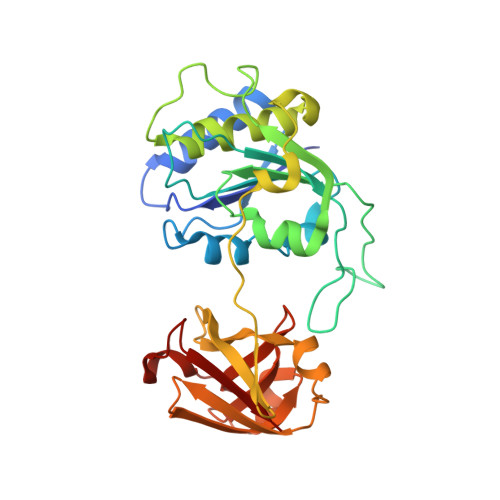
The fluorinase from Streptomyces cattleya is also a chlorinase.
Deng, H., Cobb, S.L., McEwan, A.R., McGlinchey, R.P., Naismith, J.H., O'Hagan, D., Robinson, D.A., Spencer, J.B.(2006) Angew Chem Int Ed Engl 45: 759-762
- PubMed: 16370017 Search on PubMedSearch on PubMed Central
- DOI: https://doi.org/10.1002/anie.200503582
- Primary Citation Related Structures:
2C2W - School of Chemistry and Centre for Biomolecular Science, University of St Andrews, St Andrews, KY16 9ST, UK.
Organizational Affiliation: