2BX2 | pdb_00002bx2

Catalytic domain of E. coli RNase E


Experimental Data Snapshot

  • Method: X-RAY DIFFRACTION
  • Resolution: 2.85 Å
  • R-Value Free: 
    0.257 (Depositor), 0.263 (DCC) 
  • R-Value Work: 
    0.230 (Depositor), 0.236 (DCC) 
  • R-Value Observed: 
    0.231 (Depositor) 

wwPDB Validation 3D Report Full Report

Validation slider image for 2BX2

This is version 1.2 of the entry. See complete history

Literature

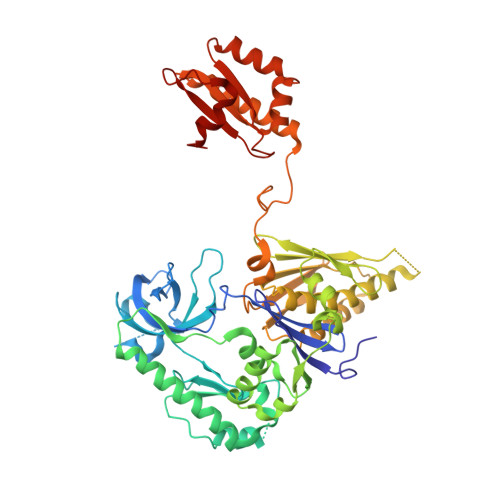
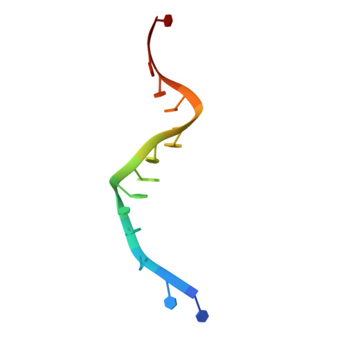
Structure of E. Coli Rnase E Catalytic Domain and Implications for RNA Processing and Turnover

Callaghan, A.J.Marcaida, M.J.Stead, J.A.Mcdowall, K.J.Scott, W.G.Luisi, B.F.

(2005) Nature 437: 1187

  • DOI: https://doi.org/10.1038/nature04084
  • Primary Citation Related Structures: 
    2BX2, 2C0B, 2C4R

  • PubMed Abstract: 

    The coordinated regulation of gene expression is required for homeostasis, growth and development in all organisms. Such coordination may be partly achieved at the level of messenger RNA stability, in which the targeted destruction of subsets of transcripts generates the potential for cross-regulating metabolic pathways. In Escherichia coli, the balance and composition of the transcript population is affected by RNase E, an essential endoribonuclease that not only turns over RNA but also processes certain key RNA precursors. RNase E cleaves RNA internally, but its catalytic power is determined by the 5' terminus of the substrate, even if this lies at a distance from the cutting site. Here we report crystal structures of the catalytic domain of RNase E as trapped allosteric intermediates with RNA substrates. Four subunits of RNase E catalytic domain associate into an interwoven quaternary structure, explaining why the subunit organization is required for catalytic activity. The subdomain encompassing the active site is structurally congruent to a deoxyribonuclease, making an unexpected link in the evolutionary history of RNA and DNA nucleases. The structure explains how the recognition of the 5' terminus of the substrate may trigger catalysis and also sheds light on the question of how RNase E might selectively process, rather than destroy, specific RNA precursors.


  • Organizational Affiliation
    • Department of Biochemistry, University of Cambridge, 80 Tennis Court Road, Cambridge CB2 1GA, UK.

Macromolecule Content 

  • Total Structure Weight: 63.02 kDa 
  • Atom Count: 4,135 
  • Modeled Residue Count: 514 
  • Deposited Residue Count: 532 
  • Unique protein chains: 1
  • Unique nucleic acid chains: 1

Macromolecules


Find similar proteins by:|  3D Structure
Entity ID: 1
MoleculeChains  Sequence LengthOrganismDetailsImage
RIBONUCLEASE EA [auth L]517Escherichia coliMutation(s): 0 
EC: 3.1.4 (PDB Primary Data), 3.1.26.12 (UniProt)
UniProt
Find proteins for P21513 (Escherichia coli (strain K12))
Explore P21513 
Go to UniProtKB:  P21513
Entity Groups
Sequence Clusters30% Identity50% Identity70% Identity90% Identity95% Identity100% Identity
UniProt GroupP21513
Sequence Annotations
Expand
Reference Sequence
Find similar nucleic acids by:  Sequence
Entity ID: 2
MoleculeChains LengthOrganismImage
RNA (5'-R(*UP*UP*UP*AP*CP*AP*GP*UP*AP*UP*UP* UP*GP*UP*U)-3')B [auth R]15N/A
Sequence Annotations
Expand
Reference Sequence

Experimental Data & Validation

Experimental Data

  • Method: X-RAY DIFFRACTION
  • Resolution: 2.85 Å
  • R-Value Free:  0.257 (Depositor), 0.263 (DCC) 
  • R-Value Work:  0.230 (Depositor), 0.236 (DCC) 
  • R-Value Observed: 0.231 (Depositor) 
Space Group: P 62 2 2
Unit Cell:
Length ( Å )Angle ( ˚ )
a = 195.838α = 90
b = 195.838β = 90
c = 143.569γ = 120
Software Package:
Software NamePurpose
REFMACrefinement
DENZOdata reduction
SCALEPACKdata scaling

Structure Validation

View Full Validation Report


Entry History 

Deposition Data

Revision History  (Full details and data files)

  • Version 1.0: 2005-10-14
    Type: Initial release
  • Version 1.1: 2011-07-13
    Changes: Advisory, Version format compliance
  • Version 1.2: 2024-05-08
    Changes: Data collection, Database references, Derived calculations, Other