2BWN | pdb_00002bwn

5-Aminolevulinate Synthase from Rhodobacter capsulatus


Experimental Data Snapshot

  • Method: X-RAY DIFFRACTION
  • Resolution: 2.10 Å
  • R-Value Free: 
    0.217 (Depositor), 0.228 (DCC) 
  • R-Value Work: 
    0.161 (Depositor), 0.172 (DCC) 
  • R-Value Observed: 
    0.164 (Depositor) 

Starting Models: experimental
View more details

wwPDB Validation 3D Report Full Report

Validation slider image for 2BWN

Ligand Structure Quality Assessment 


This is version 1.3 of the entry. See complete history

Literature

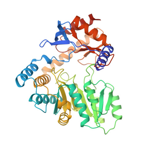
Crystal Structure of 5-Aminolevulinate Synthase, the First Enzyme of Heme Biosynthesis, and its Link to Xlsa in Humans.

Astner, I.Schulze, J.O.Van Den Heuvel, J.J.Jahn, D.Schubert, W.-D.Heinz, D.W.

(2005) EMBO J 24: 3166

  • DOI: https://doi.org/10.1038/sj.emboj.7600792
  • Primary Citation Related Structures: 
    2BWN, 2BWO, 2BWP

  • PubMed Abstract: 

    5-Aminolevulinate synthase (ALAS) is the first and rate-limiting enzyme of heme biosynthesis in humans, animals, other non-plant eukaryotes, and alpha-proteobacteria. It catalyzes the synthesis of 5-aminolevulinic acid, the first common precursor of all tetrapyrroles, from glycine and succinyl-coenzyme A (sCoA) in a pyridoxal 5'-phosphate (PLP)-dependent manner. X-linked sideroblastic anemias (XLSAs), a group of severe disorders in humans characterized by inadequate formation of heme in erythroblast mitochondria, are caused by mutations in the gene for erythroid eALAS, one of two human genes for ALAS. We present the first crystal structure of homodimeric ALAS from Rhodobacter capsulatus (ALAS(Rc)) binding its cofactor PLP. We, furthermore, present structures of ALAS(Rc) in complex with the substrates glycine or sCoA. The sequence identity of ALAS from R. capsulatus and human eALAS is 49%. XLSA-causing mutations may thus be mapped, revealing the molecular basis of XLSA in humans. Mutations are found to obstruct substrate binding, disrupt the dimer interface, or hamper the correct folding. The structure of ALAS completes the structural analysis of enzymes in heme biosynthesis.


  • Organizational Affiliation
    • Division of Structural Biology, German Research Centre for Biotechnology, Braunschweig, Germany.

Macromolecule Content 

  • Total Structure Weight: 176.37 kDa 
  • Atom Count: 13,428 
  • Modeled Residue Count: 1,586 
  • Deposited Residue Count: 1,604 
  • Unique protein chains: 1

Macromolecules

Find similar proteins by:|  3D Structure
Entity ID: 1
MoleculeChains  Sequence LengthOrganismDetailsImage
5-AMINOLEVULINATE SYNTHASEA,
B,
C [auth D],
D [auth E]
401Rhodobacter capsulatusMutation(s): 6 
EC: 2.3.1.37
UniProt
Find proteins for P18079 (Rhodobacter capsulatus (strain ATCC BAA-309 / NBRC 16581 / SB1003))
Explore P18079 
Go to UniProtKB:  P18079
Entity Groups
Sequence Clusters30% Identity50% Identity70% Identity90% Identity95% Identity100% Identity
UniProt GroupP18079
Sequence Annotations
Expand
Reference Sequence

Small Molecules

Ligands 6 Unique
IDChains Name / Formula / InChI Key2D Diagram3D Interactions
PLP

Query on PLP


Download:Ideal Coordinates CCD File
E [auth A],
H [auth B],
K [auth D],
N [auth E]
PYRIDOXAL-5'-PHOSPHATE
C8 H10 N O6 P
NGVDGCNFYWLIFO-UHFFFAOYSA-N
SIN

Query on SIN


Download:Ideal Coordinates CCD File
F [auth A],
M [auth D],
O [auth E]
SUCCINIC ACID
C4 H6 O4
KDYFGRWQOYBRFD-UHFFFAOYSA-N
SO4

Query on SO4


Download:Ideal Coordinates CCD File
I [auth B]SULFATE ION
O4 S
QAOWNCQODCNURD-UHFFFAOYSA-L
ACY

Query on ACY


Download:Ideal Coordinates CCD File
J [auth B],
L [auth D]
ACETIC ACID
C2 H4 O2
QTBSBXVTEAMEQO-UHFFFAOYSA-N
FMT

Query on FMT


Download:Ideal Coordinates CCD File
P [auth E]FORMIC ACID
C H2 O2
BDAGIHXWWSANSR-UHFFFAOYSA-N
CL

Query on CL


Download:Ideal Coordinates CCD File
G [auth A],
Q [auth E]
CHLORIDE ION
Cl
VEXZGXHMUGYJMC-UHFFFAOYSA-M

Experimental Data & Validation

Experimental Data

  • Method: X-RAY DIFFRACTION
  • Resolution: 2.10 Å
  • R-Value Free:  0.217 (Depositor), 0.228 (DCC) 
  • R-Value Work:  0.161 (Depositor), 0.172 (DCC) 
  • R-Value Observed: 0.164 (Depositor) 
Space Group: P 21 21 21
Unit Cell:
Length ( Å )Angle ( ˚ )
a = 67.875α = 90
b = 92.872β = 90
c = 250.131γ = 90
Software Package:
Software NamePurpose
REFMACrefinement
DENZOdata reduction
SCALEPACKdata scaling
EPMRphasing

Structure Validation

View Full Validation Report


Ligand Structure Quality Assessment 


Entry History 

Deposition Data

Revision History  (Full details and data files)

  • Version 1.0: 2005-09-27
    Type: Initial release
  • Version 1.1: 2011-07-13
    Changes: Advisory, Version format compliance
  • Version 1.2: 2015-12-23
    Changes: Atomic model, Derived calculations, Non-polymer description, Other, Refinement description, Structure summary
  • Version 1.3: 2023-12-13
    Changes: Data collection, Database references, Derived calculations, Other, Refinement description