2BTJ | pdb_00002btj

Fluorescent Protein EosFP - red form


Experimental Data Snapshot

  • Method: X-RAY DIFFRACTION
  • Resolution: 2.00 Å
  • R-Value Free: 
    0.232 (Depositor) 
  • R-Value Work: 
    0.198 (Depositor) 
  • R-Value Observed: 
    0.198 (Depositor) 

Starting Model: experimental
View more details

wwPDB Validation 3D Report Full Report

Validation slider image for 2BTJ

This is version 3.1 of the entry. See complete history

Literature

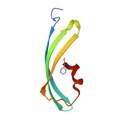
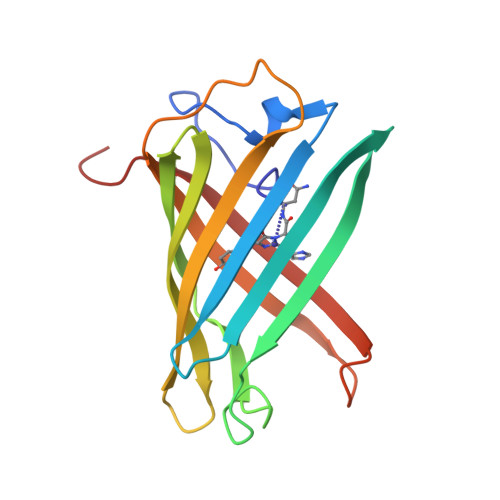
Structural Basis for Photo-Induced Protein Cleavage and Green-to-Red Conversion of Fluorescent Protein Eosfp.

Nienhaus, K.Nienhaus, G.U.Wiedenmann, J.Nar, H.

(2005) Proc Natl Acad Sci U S A 102: 9156

  • DOI: https://doi.org/10.1073/pnas.0501874102
  • Primary Citation Related Structures: 
    1ZUX, 2BTJ

  • PubMed Abstract: 

    Genetically encoded fusion constructs derived from fluorescent proteins (FPs) can be designed to report on a multitude of events and signals in cells, tissues, and entire organs without interfering with the complex machinery of life. EosFP is a novel FP from the scleractinian coral Lobophyllia hemprichii that switches its fluorescence emission from green (516 nm) to red (581 nm) upon irradiation with approximately 400-nm light. This property enables localized tagging of proteins and thus provides a valuable tool for tracking protein movements within live cells. Here, we present the x-ray structures of the green and red forms of WT EosFP. They reveal that formation of the red chromophore is associated with cleavage of the peptide backbone, with surprisingly little change elsewhere in the structure, and provide insights into the mechanism that generates this interesting posttranslational polypeptide modification.


  • Organizational Affiliation
    • Department of Biophysics and General Zoology, University of Ulm, Albert-Einstein-Allee 11, D-89081 Ulm, Germany.

Macromolecule Content 

  • Total Structure Weight: 100.82 kDa 
  • Atom Count: 7,796 
  • Modeled Residue Count: 880 
  • Deposited Residue Count: 880 
  • Unique protein chains: 2

Macromolecules

Find similar proteins by:|  3D Structure
Entity ID: 1
MoleculeChains  Sequence LengthOrganismDetailsImage
Green to red photoconvertible GFP-like protein EosFPA [auth aa],
C [auth bb],
E [auth cc],
G [auth dd]
61Lobophyllia hemprichiiMutation(s): 0 
UniProt
Find proteins for Q5S6Z9 (Lobophyllia hemprichii)
Explore Q5S6Z9 
Go to UniProtKB:  Q5S6Z9
Entity Groups
Sequence Clusters30% Identity50% Identity70% Identity90% Identity95% Identity100% Identity
UniProt GroupQ5S6Z9
Sequence Annotations
Expand
Reference Sequence
Find similar proteins by:|  3D Structure
Entity ID: 2
MoleculeChains  Sequence LengthOrganismDetailsImage
Green to red photoconvertible GFP-like protein EosFPB [auth A],
D [auth B],
F [auth C],
H [auth D]
159Lobophyllia hemprichiiMutation(s): 1 
UniProt
Find proteins for Q5S6Z9 (Lobophyllia hemprichii)
Explore Q5S6Z9 
Go to UniProtKB:  Q5S6Z9
Entity Groups
Sequence Clusters30% Identity50% Identity70% Identity90% Identity95% Identity100% Identity
UniProt GroupQ5S6Z9
Sequence Annotations
Expand
Reference Sequence

Small Molecules

Modified Residues  1 Unique
IDChains TypeFormula2D DiagramParent
NFA
Query on NFA
A [auth aa],
C [auth bb],
E [auth cc],
G [auth dd]
L-PEPTIDE LINKINGC9 H12 N2 OPHE

Experimental Data & Validation

Experimental Data

  • Method: X-RAY DIFFRACTION
  • Resolution: 2.00 Å
  • R-Value Free:  0.232 (Depositor) 
  • R-Value Work:  0.198 (Depositor) 
  • R-Value Observed: 0.198 (Depositor) 
Space Group: P 21 21 21
Unit Cell:
Length ( Å )Angle ( ˚ )
a = 70.59α = 90
b = 105.51β = 90
c = 119.62γ = 90
Software Package:
Software NamePurpose
CNSrefinement
HKLdata reduction
SCALEPACKdata scaling
PHASERphasing

Structure Validation

View Full Validation Report


Entry History 

Deposition Data

Revision History  (Full details and data files)

  • Version 1.0: 2005-06-23
    Type: Initial release
  • Version 1.1: 2013-07-24
    Changes: Atomic model, Database references, Derived calculations, Non-polymer description, Other, Refinement description, Source and taxonomy, Structure summary, Version format compliance
  • Version 1.2: 2019-10-23
    Changes: Advisory, Data collection, Database references, Derived calculations
  • Version 2.0: 2021-09-01
    Changes: Advisory, Atomic model, Data collection, Database references, Derived calculations, Non-polymer description, Other, Polymer sequence, Source and taxonomy, Structure summary
  • Version 3.0: 2023-11-15
    Changes: Atomic model, Data collection, Derived calculations
  • Version 3.1: 2023-12-13
    Changes: Refinement description