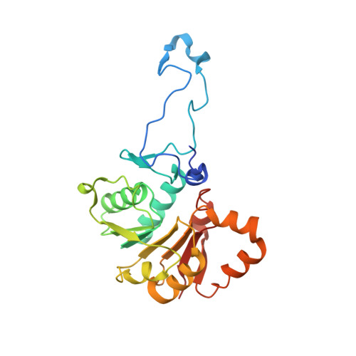
Insights Into Cephamycin Biosynthesis: The Crystal Structure of Cmci from Streptomyces Clavuligerus.
Oster, L.M., Lester, D.R., Terwisscha Van Scheltinga, A., Svenda, M., Van Lun, M., Genereux, C., Andersson, I.(2006) J Mol Biol 358: 546
- PubMed: 16527306
- DOI: https://doi.org/10.1016/j.jmb.2006.02.004
- Primary Citation of Related Structures:
2BM8, 2BM9, 2BR3, 2BR4, 2BR5 - PubMed Abstract:
Cephamycin C-producing microorganisms use two enzymes to convert cephalosporins to their 7alpha-methoxy derivatives. Here we report the X-ray structure of one of these enzymes, CmcI, from Streptomyces clavuligerus. The polypeptide chain of the enzyme folds into a C-terminal Rossmann domain and a smaller N-terminal domain, and the molecule packs as a hexamer in the crystal. The Rossmann domain binds S-adenosyl-L-methionine (SAM) and the demethylated product, S-adenosyl-L-homocysteine, in a fashion similar to the common binding mode of this cofactor in SAM-dependent methyltransferases. There is a magnesium-binding site in the vicinity of the SAM site with a bound magnesium ion ligated by residues Asp160, Glu186 and Asp187. The expected cephalosporin binding site near the magnesium ion is occupied by polyethyleneglycol (PEG) from the crystallisation medium. The geometry of the SAM and the magnesium binding sites is similar to that found in cathechol O-methyltransferase. The results suggest CmcI is a methyltransferase, and its most likely function is to catalyse the transfer of a methyl group from SAM to the 7alpha-hydroxy cephalosporin in the second catalytic reaction of cephamycin formation. Based on the docking of the putative substrate, 7alpha-hydroxy-O-carbamoyldeacetylcephalosporin C, to the structure of the ternary CmcI-Mg2+-SAM complex, we propose a model for substrate binding and catalysis. In this model, the 7-hydroxy group of the beta-lactam ring ligates the Mg2+ with its alpha-side facing the methyl group of SAM at a distance that would allow methylation of the hydroxyl-group.
Organizational Affiliation:
Department of Molecular Biology, Swedish University of Agricultural Sciences, Box 590, S-751 24 Uppsala, Sweden.


