2BMB | pdb_00002bmb

X-ray structure of the bifunctional 6-hydroxymethyl-7,8- dihydroxypterin pyrophosphokinase dihydropteroate synthase from Saccharomyces cerevisiae


Experimental Data Snapshot

  • Method: X-RAY DIFFRACTION
  • Resolution: 2.30 Å
  • R-Value Free: 
    0.216 (Depositor), 0.237 (DCC) 
  • R-Value Work: 
    0.178 (Depositor), 0.210 (DCC) 
  • R-Value Observed: 
    0.180 (Depositor) 

Starting Models: experimental
View more details

wwPDB Validation 3D Report Full Report

Validation slider image for 2BMB

Ligand Structure Quality Assessment 


This is version 1.4 of the entry. See complete history

Literature

The Three-Dimensional Structure of the Bifunctional 6-Hydroxymethyl-7,8-Dihydropterin Pyrophosphokinase/Dihydropteroate Synthase of Saccharomyces Cerevisiae

Lawrence, M.C.Iliades, P.Fernley, R.T.Berglez, J.Pilling, P.A.Macreadie, I.G.

(2005) J Mol Biology 348: 655

  • DOI: https://doi.org/10.1016/j.jmb.2005.03.021
  • Primary Citation Related Structures: 
    2BMB

  • PubMed Abstract: 

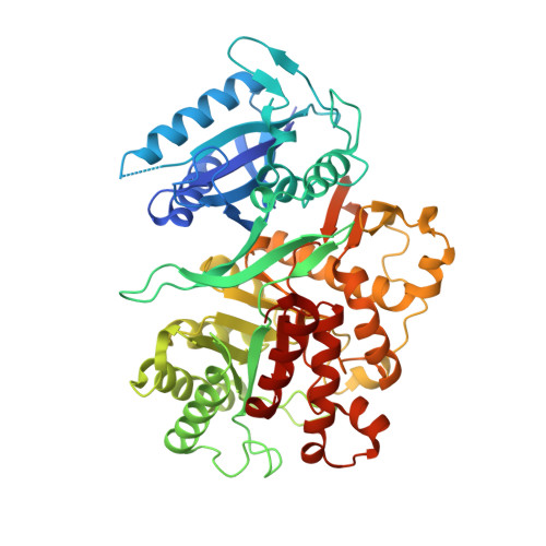
    In Saccharomyces cerevisiae and other fungi, the enzymes dihydroneopterin aldolase, 6-hydroxymethyl-7,8-dihydropterin pyrophosphokinase (HPPK) and dihydropteroate synthase (DHPS) are encoded by a polycistronic gene that is translated into a single polypeptide having all three functions. These enzymatic functions are essential to both prokaryotes and lower eukaryotes, and catalyse sequential reactions in folate biosynthesis. Deletion or disruption of either function leads to cell death. These enzymes are absent from mammals and thus make ideal antimicrobial targets. DHPS is currently the target of antifolate therapy for a number of infectious diseases, and its activity is inhibited by sulfonamides and sulfones. These drugs are typically used as part of a synergistic cocktail with the 2,4-diaminopyrimidines that inhibit dihydrofolate reductase. A gene encoding the S.cerevisiae HPPK and DHPS enzymes has been cloned and expressed in Escherichia coli. A complex of the purified bifunctional polypeptide with a pterin monophosphate substrate analogue has been crystallized, and its structure solved by molecular replacement and refined to 2.3A resolution. The polypeptide consists of two structural domains, each of which closely resembles its respective monofunctional bacterial HPPK and DHPS counterpart. The mode of ligand binding is similar to that observed in the bacterial enzymes. The association between the domains within the polypeptide as well as the quaternary association of the polypeptide via its constituent DHPS domains provide insight into the assembly of the trifunctional enzyme in S.cerevisiae and probably other fungal species.


  • Organizational Affiliation
    • CSIRO Health Sciences and Nutrition, 343 Royal Parade, Parkville, Victoria 3052, Australia. mike.lawrence@csiro.au

Macromolecule Content 

  • Total Structure Weight: 62.25 kDa 
  • Atom Count: 4,282 
  • Modeled Residue Count: 513 
  • Deposited Residue Count: 545 
  • Unique protein chains: 1

Macromolecules

Find similar proteins by:|  3D Structure
Entity ID: 1
MoleculeChains  Sequence LengthOrganismDetailsImage
FOLIC ACID SYNTHESIS PROTEIN FOL1545Saccharomyces cerevisiaeMutation(s): 0 
EC: 2.7.6.3 (PDB Primary Data), 6.3.2.12 (PDB Primary Data), 2.5.1.15 (UniProt), 4.1.2.25 (UniProt)
Membrane Entity: Yes 
UniProt
Find proteins for P53848 (Saccharomyces cerevisiae (strain ATCC 204508 / S288c))
Explore P53848 
Go to UniProtKB:  P53848
Entity Groups
Sequence Clusters30% Identity50% Identity70% Identity90% Identity95% Identity100% Identity
UniProt GroupP53848
Sequence Annotations
Expand
Reference Sequence

Experimental Data & Validation

Experimental Data

  • Method: X-RAY DIFFRACTION
  • Resolution: 2.30 Å
  • R-Value Free:  0.216 (Depositor), 0.237 (DCC) 
  • R-Value Work:  0.178 (Depositor), 0.210 (DCC) 
  • R-Value Observed: 0.180 (Depositor) 
Space Group: P 41 21 2
Unit Cell:
Length ( Å )Angle ( ˚ )
a = 92.566α = 90
b = 92.566β = 90
c = 192.401γ = 90
Software Package:
Software NamePurpose
REFMACrefinement
DENZOdata reduction
SCALEPACKdata scaling
MOLREPphasing

Structure Validation

View Full Validation Report


Ligand Structure Quality Assessment 


Entry History 

Deposition Data

Revision History  (Full details and data files)

  • Version 1.0: 2006-01-17
    Type: Initial release
  • Version 1.1: 2011-05-08
    Changes: Version format compliance
  • Version 1.2: 2011-07-13
    Changes: Version format compliance
  • Version 1.3: 2017-07-05
    Changes: Data collection
  • Version 1.4: 2023-12-13
    Changes: Data collection, Database references, Other, Refinement description