2BLF

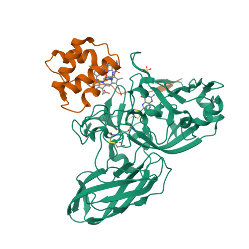
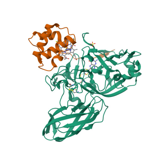
Sulfite dehydrogenase from Starkeya Novella


Experimental Data Snapshot

  • Method: X-RAY DIFFRACTION
  • Resolution: 1.80 Å
  • R-Value Free: 0.169 
  • R-Value Work: 0.136 
  • R-Value Observed: 0.138 

Starting Model: experimental
View more details

wwPDB Validation   3D Report Full Report


Ligand Structure Quality Assessment 

Created with Raphaël 2.3.0Worse 01 BetterLigand structure goodness of fit to experimental dataBest fitted HECClick on this verticalbar to view detailsBest fitted MSSClick on this verticalbar to view details

This is version 2.2 of the entry. See complete history


Literature

Molecular Basis of Intramolecular Electron Transfer in Sulfite-Oxidizing Enzymes is Revealed by High Resolution Structure of a Heterodimeric Complex of the Catalytic Molybdopterin Subunit and a C-Type Cytochrome Subunit.

Kappler, U.Bailey, S.

(2005) J Biol Chem 280: 24999

  • DOI: https://doi.org/10.1074/jbc.M503237200
  • Primary Citation of Related Structures:  
    2BLF, 2BPB

  • PubMed Abstract: 

    Sulfite-oxidizing molybdoenzymes convert the highly reactive and therefore toxic sulfite to sulfate and have been identified in insects, animals, plants, and bacteria. Although the well studied enzymes from higher animals serve to detoxify sulfite that arises from the catabolism of sulfur-containing amino acids, the bacterial enzymes have a central role in converting sulfite formed during dissimilatory oxidation of reduced sulfur compounds. Here we describe the structure of the Starkeya novella sulfite dehydrogenase, a heterodimeric complex of the catalytic molybdopterin subunit and a c-type cytochrome subunit, that reveals the molecular mechanism of intramolecular electron transfer in sulfite-oxidizing enzymes. The close approach of the two redox centers in the protein complex (Mo-Fe distance 16.6 A) allows for rapid electron transfer via tunnelling or aided by the protein environment. The high resolution structure of the complex has allowed the identification of potential through-bond pathways for electron transfer including a direct link via Arg-55A and/or an aromatic-mediated pathway. A potential site of electron transfer to an external acceptor cytochrome c was also identified on the SorB subunit on the opposite side to the interaction with the catalytic SorA subunit.


  • Organizational Affiliation

    Centre for Metals in Biology, University of Queensland, St. Lucia, Queensland 4072, Australia.


Macromolecules
Find similar proteins by:  (by identity cutoff)  |  3D Structure
Entity ID: 1
MoleculeChains Sequence LengthOrganismDetailsImage
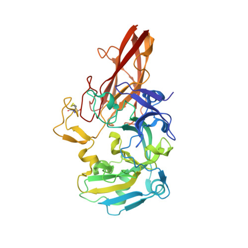
Sulfite:cytochrome c oxidoreductase subunit A373Ancylobacter novellusMutation(s): 0 
Gene Names: sorA
UniProt
Find proteins for Q9LA16 (Ancylobacter novellus)
Explore Q9LA16 
Go to UniProtKB:  Q9LA16
Entity Groups  
Sequence Clusters30% Identity50% Identity70% Identity90% Identity95% Identity100% Identity
UniProt GroupQ9LA16
Sequence Annotations
Expand
  • Reference Sequence
Find similar proteins by:  (by identity cutoff)  |  3D Structure
Entity ID: 2
MoleculeChains Sequence LengthOrganismDetailsImage
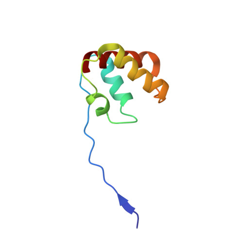
Sulfite:cytochrome c oxidoreductase subunit B81Ancylobacter novellusMutation(s): 0 
Gene Names: sorB
UniProt
Find proteins for Q9LA15 (Ancylobacter novellus)
Explore Q9LA15 
Go to UniProtKB:  Q9LA15
Entity Groups  
Sequence Clusters30% Identity50% Identity70% Identity90% Identity95% Identity100% Identity
UniProt GroupQ9LA15
Sequence Annotations
Expand
  • Reference Sequence
Experimental Data & Validation

Experimental Data

  • Method: X-RAY DIFFRACTION
  • Resolution: 1.80 Å
  • R-Value Free: 0.169 
  • R-Value Work: 0.136 
  • R-Value Observed: 0.138 
  • Space Group: P 21 21 2
Unit Cell:
Length ( Å )Angle ( ˚ )
a = 97.466α = 90
b = 92.513β = 90
c = 55.897γ = 90
Software Package:
Software NamePurpose
REFMACrefinement
MOSFLMdata reduction
SCALAdata scaling
CCP4phasing

Structure Validation

View Full Validation Report


Ligand Structure Quality Assessment 

Created with Raphaël 2.3.0Worse 01 BetterLigand structure goodness of fit to experimental dataBest fitted HECClick on this verticalbar to view detailsBest fitted MSSClick on this verticalbar to view details

Entry History 

Deposition Data

Revision History  (Full details and data files)

  • Version 1.0: 2005-04-28
    Type: Initial release
  • Version 1.1: 2014-10-22
    Changes: Database references, Derived calculations, Non-polymer description, Other, Structure summary, Version format compliance
  • Version 2.0: 2018-02-07
    Changes: Advisory, Atomic model, Database references, Derived calculations, Source and taxonomy, Structure summary
  • Version 2.1: 2023-12-13
    Changes: Data collection, Database references, Derived calculations, Other, Refinement description
  • Version 2.2: 2024-11-20
    Changes: Structure summary