2BKJ

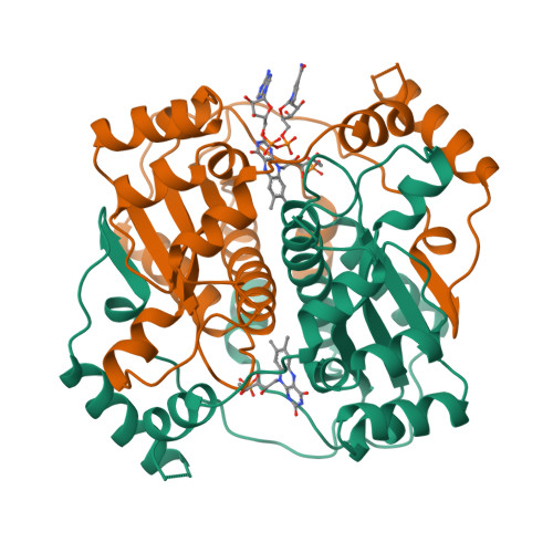
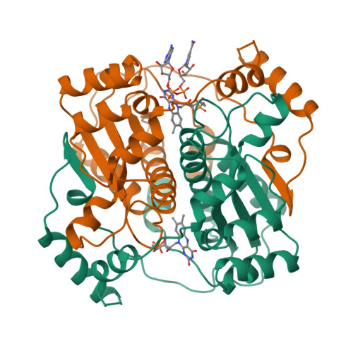
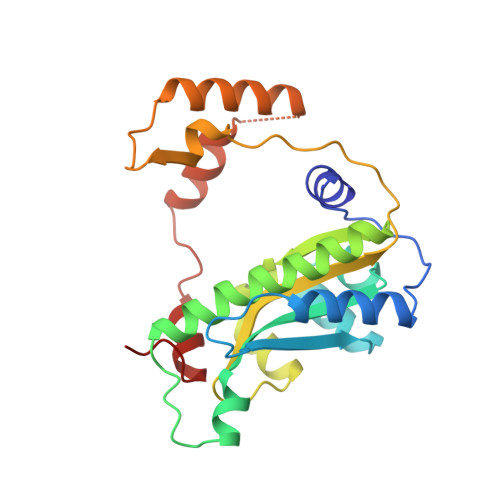
NADPH:FMN OXIDOREDUCTASE FROM VIBRIO HARVEYI COMPLEXED WITH NAD+


Experimental Data Snapshot

  • Method: X-RAY DIFFRACTION
  • Resolution: 2.08 Å
  • R-Value Free: 0.206 
  • R-Value Work: 0.185 
  • R-Value Observed: 0.185 

Starting Model: experimental
View more details

wwPDB Validation   3D Report Full Report


Ligand Structure Quality Assessment 


This is version 1.5 of the entry. See complete history


Literature

Unusual folded conformation of nicotinamide adenine dinucleotide bound to flavin reductase P.

Tanner, J.J.Tu, S.C.Barbour, L.J.Barnes, C.L.Krause, K.L.

(1999) Protein Sci 8: 1725-1732

  • DOI: https://doi.org/10.1110/ps.8.9.1725
  • Primary Citation of Related Structures:  
    2BKJ

  • PubMed Abstract: 

    The 2.1 A resolution crystal structure of flavin reductase P with the inhibitor nicotinamide adenine dinucleotide (NAD) bound in the active site has been determined. NAD adopts a novel, folded conformation in which the nicotinamide and adenine rings stack in parallel with an inter-ring distance of 3.6 A. The pyrophosphate binds next to the flavin cofactor isoalloxazine, while the stacked nicotinamide/adenine moiety faces away from the flavin. The observed NAD conformation is quite different from the extended conformations observed in other enzyme/NAD(P) structures; however, it resembles the conformation proposed for NAD in solution. The flavin reductase P/NAD structure provides new information about the conformational diversity of NAD, which is important for understanding catalysis. This structure offers the first crystallographic evidence of a folded NAD with ring stacking, and it is the first enzyme structure containing an FMN cofactor interacting with NAD(P). Analysis of the structure suggests a possible dynamic mechanism underlying NADPH substrate specificity and product release that involves unfolding and folding of NADP(H).


  • Organizational Affiliation

    Department of Chemistry, University of Missouri-Columbia 65211, USA.


Macromolecules
Find similar proteins by:  (by identity cutoff)  |  3D Structure
Entity ID: 1
MoleculeChains Sequence LengthOrganismDetailsImage
FLAVIN REDUCTASE
A, B
240Vibrio harveyiMutation(s): 0 
EC: 1.6.99 (PDB Primary Data), 1.5.1.38 (UniProt)
UniProt
Find proteins for Q56691 (Vibrio harveyi)
Explore Q56691 
Go to UniProtKB:  Q56691
Entity Groups  
Sequence Clusters30% Identity50% Identity70% Identity90% Identity95% Identity100% Identity
UniProt GroupQ56691
Sequence Annotations
Expand
  • Reference Sequence
Small Molecules
Ligands 2 Unique
IDChains Name / Formula / InChI Key2D Diagram3D Interactions
NAD
Query on NAD

Download Ideal Coordinates CCD File 
E [auth B]NICOTINAMIDE-ADENINE-DINUCLEOTIDE
C21 H27 N7 O14 P2
BAWFJGJZGIEFAR-NNYOXOHSSA-N
FMN
Query on FMN

Download Ideal Coordinates CCD File 
C [auth A],
D [auth B]
FLAVIN MONONUCLEOTIDE
C17 H21 N4 O9 P
FVTCRASFADXXNN-SCRDCRAPSA-N
Experimental Data & Validation

Experimental Data

  • Method: X-RAY DIFFRACTION
  • Resolution: 2.08 Å
  • R-Value Free: 0.206 
  • R-Value Work: 0.185 
  • R-Value Observed: 0.185 
  • Space Group: P 1 21 1
Unit Cell:
Length ( Å )Angle ( ˚ )
a = 51.2α = 90
b = 85.9β = 114.7
c = 59.2γ = 90
Software Package:
Software NamePurpose
AMoREphasing
X-PLORrefinement
MADNESdata reduction
XSCALEdata scaling

Structure Validation

View Full Validation Report


Ligand Structure Quality Assessment 


Entry History 

Deposition Data

Revision History  (Full details and data files)

  • Version 1.0: 1999-09-10
    Type: Initial release
  • Version 1.1: 2008-03-24
    Changes: Version format compliance
  • Version 1.2: 2011-07-13
    Changes: Version format compliance
  • Version 1.3: 2018-04-18
    Changes: Data collection
  • Version 1.4: 2023-08-09
    Changes: Database references, Derived calculations, Refinement description
  • Version 1.5: 2024-05-22
    Changes: Data collection