2BJW | pdb_00002bjw

PspF AAA domain


Experimental Data Snapshot

  • Method: X-RAY DIFFRACTION
  • Resolution: 1.75 Å
  • R-Value Free: 
    0.208 (Depositor), 0.225 (DCC) 
  • R-Value Work: 
    0.180 (Depositor), 0.197 (DCC) 
  • R-Value Observed: 
    0.180 (Depositor) 

Starting Model: experimental
View more details

wwPDB Validation 3D Report Full Report

Validation slider image for 2BJW

This is version 1.3 of the entry. See complete history

Literature

Structural Insights Into the Activity of Enhancer-Binding Proteins

Rappas, M.Schumacher, J.Beuron, F.Niwa, H.Bordes, P.Wigneshweraraj, S.Keetch, C.A.Robinson, C.V.Buck, M.Zhang, X.

(2005) Science 307: 1972

  • DOI: https://doi.org/10.1126/science.1105932
  • Primary Citation Related Structures: 
    2BJV, 2BJW

  • PubMed Abstract: 

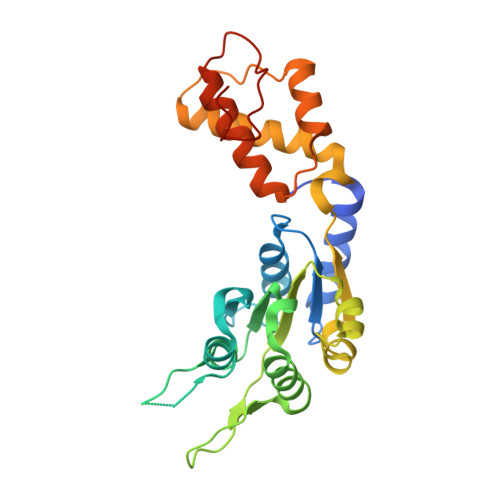
    Activators of bacterial sigma54-RNA polymerase holoenzyme are mechanochemical proteins that use adenosine triphosphate (ATP) hydrolysis to activate transcription. We have determined by cryogenic electron microscopy (cryo-EM) a 20 angstrom resolution structure of an activator, phage shock protein F [PspF(1-275)], which is bound to an ATP transition state analog in complex with its basal factor, sigma54. By fitting the crystal structure of PspF(1-275) at 1.75 angstroms into the EM map, we identified two loops involved in binding sigma54. Comparing enhancer-binding structures in different nucleotide states and mutational analysis led us to propose nucleotide-dependent conformational changes that free the loops for association with sigma54.


  • Organizational Affiliation
    • Department of Biological Sciences, Imperial College London, London, SW7 2AZ, UK.

Macromolecule Content 

  • Total Structure Weight: 29.99 kDa 
  • Atom Count: 2,113 
  • Modeled Residue Count: 243 
  • Deposited Residue Count: 265 
  • Unique protein chains: 1

Macromolecules

Find similar proteins by:|  3D Structure
Entity ID: 1
MoleculeChains  Sequence LengthOrganismDetailsImage
PSP OPERON TRANSCRIPTIONAL ACTIVATOR265Escherichia coli K-12Mutation(s): 0 
UniProt
Find proteins for P37344 (Escherichia coli (strain K12))
Explore P37344 
Go to UniProtKB:  P37344
Entity Groups
Sequence Clusters30% Identity50% Identity70% Identity90% Identity95% Identity100% Identity
UniProt GroupP37344
Sequence Annotations
Expand
Reference Sequence

Experimental Data & Validation

Experimental Data

  • Method: X-RAY DIFFRACTION
  • Resolution: 1.75 Å
  • R-Value Free:  0.208 (Depositor), 0.225 (DCC) 
  • R-Value Work:  0.180 (Depositor), 0.197 (DCC) 
  • R-Value Observed: 0.180 (Depositor) 
Space Group: P 65
Unit Cell:
Length ( Å )Angle ( ˚ )
a = 113.695α = 90
b = 113.695β = 90
c = 39.277γ = 120
Software Package:
Software NamePurpose
CNSrefinement
DENZOdata reduction
SCALEPACKdata scaling
PHASERphasing

Structure Validation

View Full Validation Report


Entry History 

Deposition Data

Revision History  (Full details and data files)

  • Version 1.0: 2005-03-31
    Type: Initial release
  • Version 1.1: 2011-05-08
    Changes: Version format compliance
  • Version 1.2: 2011-07-13
    Changes: Version format compliance
  • Version 1.3: 2023-12-13
    Changes: Data collection, Database references, Other, Refinement description