2BIT | pdb_00002bit

Crystal structure of human cyclophilin D at 1.7 A resolution


Experimental Data Snapshot

  • Method: X-RAY DIFFRACTION
  • Resolution: 1.71 Å
  • R-Value Free: 
    0.175 (Depositor), 0.176 (DCC) 
  • R-Value Work: 
    0.139 (Depositor), 0.143 (DCC) 
  • R-Value Observed: 
    0.141 (Depositor) 

Starting Model: experimental
View more details

wwPDB Validation 3D Report Full Report

Validation slider image for 2BIT

This is version 1.4 of the entry. See complete history

Literature

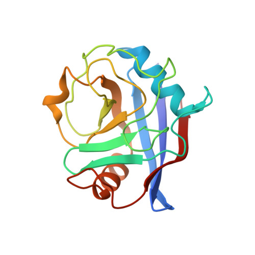
Crystal Engineering Yields Crystals of Cyclophilin D Diffracting to 1.7 A Resolution

Schlatter, D.Thoma, R.Kueng, E.Stihle, M.Mueller, F.Boroni, E.Hennig, M.

(2005) Acta Crystallogr D Biol Crystallogr 61: 513-519

  • DOI: https://doi.org/10.1107/S0907444905003070
  • Primary Citation Related Structures: 
    2BIT, 2BIU

  • PubMed Abstract: 

    In the pharmaceutical industry, knowledge of the three-dimensional structure of a specific target facilitates the drug-discovery process. Despite possessing favoured analytical properties such as high purity and monodispersion in light scattering, some proteins are not capable of forming crystals suitable for X-ray analysis. Cyclophilin D, an isoform of cyclophilin that is expressed in the mitochondria, was selected as a drug target for the treatment of cardiac disorders. As the wild-type enzyme defied all attempts at crystallization, protein engineering on the enzyme surface was performed. The K133I mutant gave crystals that diffracted to 1.7 A resolution using in-house X-ray facilities and were suitable for soaking experiments. The crystals were very robust and diffraction was maintained after soaking in 25% DMSO solution: excellent conditions for the rapid analysis of complex structures including crystallographic fragment screening.


  • Organizational Affiliation
    • F. Hoffmann-La Roche Ltd, Pharmaceutical Research Discovery, CH-4070 Basel, Switzerland. daniel.schlatter@roche.com

Macromolecule Content 

  • Total Structure Weight: 17.74 kDa 
  • Atom Count: 1,633 
  • Modeled Residue Count: 164 
  • Deposited Residue Count: 165 
  • Unique protein chains: 1

Macromolecules

Find similar proteins by:|  3D Structure
Entity ID: 1
MoleculeChains  Sequence LengthOrganismDetailsImage
PEPTIDYL-PROLYL CIS-TRANS ISOMERASEA [auth X]165Homo sapiensMutation(s): 1 
EC: 5.2.1.8
UniProt & NIH Common Fund Data Resources
Find proteins for P30405 (Homo sapiens)
Explore P30405 
Go to UniProtKB:  P30405
PHAROS:  P30405
GTEx:  ENSG00000108179 
Entity Groups
Sequence Clusters30% Identity50% Identity70% Identity90% Identity95% Identity100% Identity
UniProt GroupP30405
Sequence Annotations
Expand
Reference Sequence

Experimental Data & Validation

Experimental Data

  • Method: X-RAY DIFFRACTION
  • Resolution: 1.71 Å
  • R-Value Free:  0.175 (Depositor), 0.176 (DCC) 
  • R-Value Work:  0.139 (Depositor), 0.143 (DCC) 
  • R-Value Observed: 0.141 (Depositor) 
Space Group: P 41 21 2
Unit Cell:
Length ( Å )Angle ( ˚ )
a = 57.165α = 90
b = 57.165β = 90
c = 87.227γ = 90
Software Package:
Software NamePurpose
REFMACrefinement
DENZOdata reduction
SCALEPACKdata scaling
AMoREphasing

Structure Validation

View Full Validation Report


Entry History 

Deposition Data

Revision History  (Full details and data files)

  • Version 1.0: 2005-01-26
    Type: Initial release
  • Version 1.1: 2011-05-08
    Changes: Version format compliance
  • Version 1.2: 2011-07-13
    Changes: Version format compliance
  • Version 1.3: 2018-06-13
    Changes: Data collection, Database references
  • Version 1.4: 2023-12-13
    Changes: Data collection, Database references, Other, Refinement description