2BGT | pdb_00002bgt

CRYSTAL STRUCTURE OF THE DNA MODIFYING ENZYME BETA-GLUCOSYLTRANSFERASE IN THE PRESENCE AND ABSENCE OF THE SUBSTRATE URIDINE DIPHOSPHOGLUCOSE


Experimental Data Snapshot

  • Method: X-RAY DIFFRACTION
  • Resolution: 2.20 Å
  • R-Value Work: 
    0.194 (Depositor) 
  • R-Value Observed: 
    0.194 (Depositor) 

wwPDB Validation 3D Report Full Report

Validation slider image for 2BGT

This is version 1.4 of the entry. See complete history

Literature

Crystal structure of the DNA modifying enzyme beta-glucosyltransferase in the presence and absence of the substrate uridine diphosphoglucose.

Vrielink, A.Ruger, W.Driessen, H.P.Freemont, P.S.

(1994) EMBO J 13: 3413-3422

  • DOI: https://doi.org/10.1002/j.1460-2075.1994.tb06646.x
  • Primary Citation Related Structures: 
    1BGT, 1BGU, 2BGT, 2BGU

  • PubMed Abstract: 

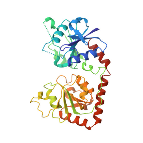
    Bacteriophage T4 beta-glucosyltransferase (EC 2.4.1.27) catalyses the transfer of glucose from uridine diphosphoglucose to hydroxymethyl groups of modified cytosine bases in T4 duplex DNA forming beta-glycosidic linkages. The enzyme forms part of a phage DNA protection system. We have solved and refined the crystal structure of recombinant beta-glucosyltransferase to 2.2 A resolution in the presence and absence of the substrate, uridine diphosphoglucose. The structure comprises two domains of similar topology, each reminiscent of a nucleotide binding fold. The two domains are separated by a central cleft which generates a concave surface along one side of the molecule. The substrate-bound complex reveals only clear electron density for the uridine diphosphate portion of the substrate. The UDPG is bound in a pocket at the bottom of the cleft between the two domains and makes extensive hydrogen bonding contacts with residues of the C-terminal domain only. The domains undergo a rigid body conformational change causing the structure to adopt a more closed conformation upon ligand binding. The movement of the domains is facilitated by a hinge region between residues 166 and 172. Electrostatic surface potential calculations reveal a large positive potential along the concave surface of the structure, suggesting a possible site for duplex DNA interaction.


  • Organizational Affiliation
    • Protein Structure Laboratory, Imperial Cancer Research Fund, London.

Macromolecule Content 

  • Total Structure Weight: 40.72 kDa 
  • Atom Count: 2,867 
  • Modeled Residue Count: 329 
  • Deposited Residue Count: 351 
  • Unique protein chains: 1

Macromolecules

Find similar proteins by:|  3D Structure
Entity ID: 1
MoleculeChains  Sequence LengthOrganismDetailsImage
BETA-GLUCOSYLTRANSFERASE351Tequatrovirus T4Mutation(s): 0 
EC: 2.4.1.27
UniProt
Find proteins for P04547 (Enterobacteria phage T4)
Explore P04547 
Go to UniProtKB:  P04547
Entity Groups
Sequence Clusters30% Identity50% Identity70% Identity90% Identity95% Identity100% Identity
UniProt GroupP04547
Sequence Annotations
Expand
Reference Sequence

Experimental Data & Validation

Experimental Data

  • Method: X-RAY DIFFRACTION
  • Resolution: 2.20 Å
  • R-Value Work:  0.194 (Depositor) 
  • R-Value Observed: 0.194 (Depositor) 
Space Group: P 21 21 2
Unit Cell:
Length ( Å )Angle ( ˚ )
a = 152.88α = 90
b = 52.25β = 90
c = 53.66γ = 90
Software Package:
Software NamePurpose
X-PLORmodel building
X-PLORrefinement
X-PLORphasing

Structure Validation

View Full Validation Report


Entry History 

Deposition Data

Revision History  (Full details and data files)

  • Version 1.0: 1995-12-09
    Type: Initial release
  • Version 1.1: 2008-03-24
    Changes: Version format compliance
  • Version 1.2: 2011-07-13
    Changes: Version format compliance
  • Version 1.3: 2017-11-29
    Changes: Advisory, Derived calculations, Other
  • Version 1.4: 2024-02-14
    Changes: Advisory, Data collection, Database references