2BF8 | pdb_00002bf8

Crystal structure of SUMO modified ubiquitin conjugating enzyme E2- 25K


Experimental Data Snapshot

  • Method: X-RAY DIFFRACTION
  • Resolution: 2.30 Å
  • R-Value Free: 
    0.279 (Depositor) 
  • R-Value Work: 
    0.210 (Depositor), 0.213 (DCC) 
  • R-Value Observed: 
    0.213 (Depositor) 

Starting Model: experimental
View more details

wwPDB Validation 3D Report Full Report

Validation slider image for 2BF8

This is version 1.2 of the entry. See complete history

Literature

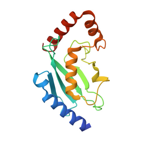
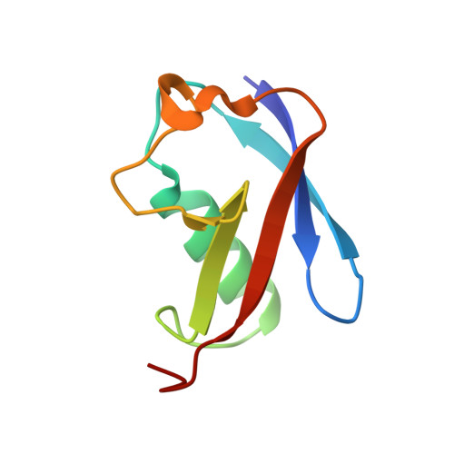
Sumo Imodification of the Ubiquitin Conjugating Enzyme E2-25K

Pichler, A.Knipscheer, P.Oberhofer, E.Van Dijk, W.J.Korner, R.Velgaard Olsen, J.Jentsch, S.Melchior, F.Sixma, T.K.

(2005) Nat Struct Mol Biol 12: 264

  • DOI: https://doi.org/10.1038/nsmb903
  • Primary Citation Related Structures: 
    2BEP, 2BF8

  • PubMed Abstract: 

    Post-translational modification with small ubiquitin-related modifier (SUMO) alters the function of many proteins, but the molecular mechanisms and consequences of this modification are still poorly defined. During a screen for novel SUMO1 targets, we identified the ubiquitin-conjugating enzyme E2-25K (Hip2). SUMO attachment severely impairs E2-25K ubiquitin thioester and unanchored ubiquitin chain formation in vitro. Crystal structures of E2-25K(1-155) and of the E2-25K(1-155)-SUMO conjugate (E2-25K(*)SUMO) indicate that SUMO attachment interferes with E1 interaction through its location on the N-terminal helix. The SUMO acceptor site in E2-25K, Lys14, does not conform to the consensus site found in most SUMO targets (PsiKXE), and functions only in the context of an alpha-helix. In contrast, adjacent SUMO consensus sites are modified only when in unstructured peptides. The demonstration that secondary structure elements are part of SUMO attachment signals could contribute to a better prediction of SUMO targets.


  • Organizational Affiliation
    • University of Göttingen, Department of Biochemistry I, Humboldt Allee 23, 37073 Göttingen, Germany.

Macromolecule Content 

  • Total Structure Weight: 26.82 kDa 
  • Atom Count: 1,947 
  • Modeled Residue Count: 231 
  • Deposited Residue Count: 236 
  • Unique protein chains: 2

Macromolecules

Find similar proteins by:|  3D Structure
Entity ID: 1
MoleculeChains  Sequence LengthOrganismDetailsImage
UBIQUITIN-CONJUGATING ENZYME E2-25 KDA159Bos taurusMutation(s): 0 
EC: 6.3.2.19 (PDB Primary Data), 2.3.2.23 (UniProt)
UniProt
Find proteins for P61085 (Bos taurus)
Explore P61085 
Go to UniProtKB:  P61085
Entity Groups
Sequence Clusters30% Identity50% Identity70% Identity90% Identity95% Identity100% Identity
UniProt GroupP61085
Sequence Annotations
Expand
Reference Sequence
Find similar proteins by:|  3D Structure
Entity ID: 2
MoleculeChains  Sequence LengthOrganismDetailsImage
UBIQUITIN-LIKE PROTEIN SMT3C77Homo sapiensMutation(s): 0 
UniProt & NIH Common Fund Data Resources
Find proteins for P63165 (Homo sapiens)
Explore P63165 
Go to UniProtKB:  P63165
PHAROS:  P63165
GTEx:  ENSG00000116030 
Entity Groups
Sequence Clusters30% Identity50% Identity70% Identity90% Identity95% Identity100% Identity
UniProt GroupP63165
Sequence Annotations
Expand
Reference Sequence

Experimental Data & Validation

Experimental Data

  • Method: X-RAY DIFFRACTION
  • Resolution: 2.30 Å
  • R-Value Free:  0.279 (Depositor) 
  • R-Value Work:  0.210 (Depositor), 0.213 (DCC) 
  • R-Value Observed: 0.213 (Depositor) 
Space Group: P 43 21 2
Unit Cell:
Length ( Å )Angle ( ˚ )
a = 58.391α = 90
b = 58.391β = 90
c = 162.842γ = 90
Software Package:
Software NamePurpose
REFMACrefinement
MOSFLMdata reduction
SCALAdata scaling
AMoREphasing

Structure Validation

View Full Validation Report


Entry History 

Deposition Data

Revision History  (Full details and data files)

  • Version 1.0: 2005-02-16
    Type: Initial release
  • Version 1.1: 2011-07-13
    Changes: Advisory, Version format compliance
  • Version 1.2: 2023-12-13
    Changes: Data collection, Database references, Other, Refinement description