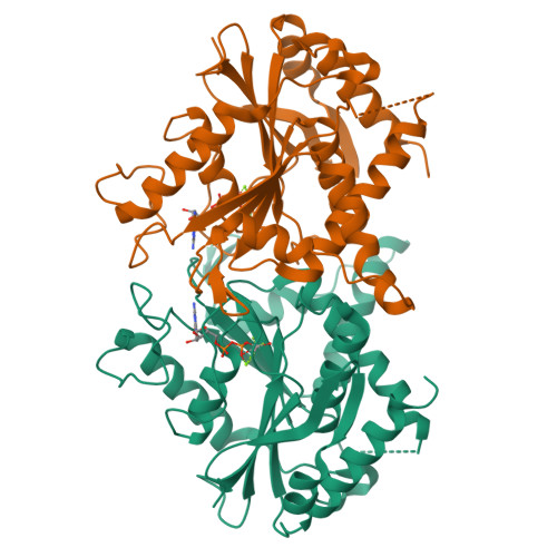
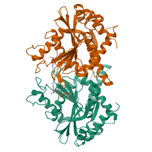
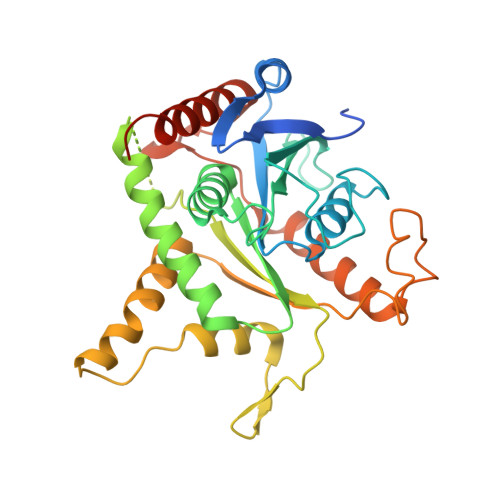
2B92

Crystal-structure of the N-terminal Large GTPase Domain of human Guanylate Binding protein 1 (hGBP1) in complex with GDP/AlF3


Experimental Data Snapshot

  • Method: X-RAY DIFFRACTION
  • Resolution: 3.20 Å
  • R-Value Free: 0.250 
  • R-Value Work: 0.204 
  • R-Value Observed: 0.207 

Starting Model: experimental
View more details

wwPDB Validation   3D Report Full Report


Ligand Structure Quality Assessment 


This is version 1.4 of the entry. See complete history


Literature

How guanylate-binding proteins achieve assembly-stimulated processive cleavage of GTP to GMP.

Ghosh, A.Praefcke, G.J.Renault, L.Wittinghofer, A.Herrmann, C.

(2006) Nature 440: 101-104

  • DOI: https://doi.org/10.1038/nature04510
  • Primary Citation of Related Structures:  
    2B8W, 2B92, 2BC9, 2D4H

  • PubMed Abstract: 

    Interferons are immunomodulatory cytokines that mediate anti-pathogenic and anti-proliferative effects in cells. Interferon-gamma-inducible human guanylate binding protein 1 (hGBP1) belongs to the family of dynamin-related large GTP-binding proteins, which share biochemical properties not found in other families of GTP-binding proteins such as nucleotide-dependent oligomerization and fast cooperative GTPase activity. hGBP1 has an additional property by which it hydrolyses GTP to GMP in two consecutive cleavage reactions. Here we show that the isolated amino-terminal G domain of hGBP1 retains the main enzymatic properties of the full-length protein and can cleave GDP directly. Crystal structures of the N-terminal G domain trapped at successive steps along the reaction pathway and biochemical data reveal the molecular basis for nucleotide-dependent homodimerization and cleavage of GTP. Similar to effector binding in other GTP-binding proteins, homodimerization is regulated by structural changes in the switch regions. Homodimerization generates a conformation in which an arginine finger and a serine are oriented for efficient catalysis. Positioning of the substrate for the second hydrolysis step is achieved by a change in nucleotide conformation at the ribose that keeps the guanine base interactions intact and positions the beta-phosphates in the gamma-phosphate-binding site.


  • Organizational Affiliation

    Abteilung Strukturelle Biologie, Max-Planck-Institut für Molekulare Physiologie, Otto-Hahn-Strasse 11, 44227 Dortmund, Germany.


Macromolecules
Find similar proteins by:  (by identity cutoff)  |  3D Structure
Entity ID: 1
MoleculeChains Sequence LengthOrganismDetailsImage
Interferon-induced guanylate-binding protein 1
A, B
328Homo sapiensMutation(s): 0 
Gene Names: GBP1
EC: 3.6.1 (UniProt), 3.6.5 (UniProt)
UniProt & NIH Common Fund Data Resources
Find proteins for P32455 (Homo sapiens)
Explore P32455 
Go to UniProtKB:  P32455
PHAROS:  P32455
GTEx:  ENSG00000117228 
Entity Groups  
Sequence Clusters30% Identity50% Identity70% Identity90% Identity95% Identity100% Identity
UniProt GroupP32455
Sequence Annotations
Expand
  • Reference Sequence
Experimental Data & Validation

Experimental Data

  • Method: X-RAY DIFFRACTION
  • Resolution: 3.20 Å
  • R-Value Free: 0.250 
  • R-Value Work: 0.204 
  • R-Value Observed: 0.207 
  • Space Group: P 21 21 21
Unit Cell:
Length ( Å )Angle ( ˚ )
a = 148.82α = 90
b = 103.921β = 90
c = 55.485γ = 90
Software Package:
Software NamePurpose
REFMACrefinement
MAR345data collection
XDSdata scaling
AMoREphasing

Structure Validation

View Full Validation Report


Ligand Structure Quality Assessment 


Entry History 

Deposition Data

Revision History  (Full details and data files)

  • Version 1.0: 2006-03-07
    Type: Initial release
  • Version 1.1: 2008-05-01
    Changes: Version format compliance
  • Version 1.2: 2011-07-13
    Changes: Advisory, Version format compliance
  • Version 1.3: 2017-10-11
    Changes: Refinement description
  • Version 1.4: 2023-08-23
    Changes: Data collection, Database references, Derived calculations, Refinement description