2B8J | pdb_00002b8j

Crystal structure of AphA class B acid phosphatase/phosphotransferase ternary complex with adenosine and phosphate at 2 A resolution


Experimental Data Snapshot

  • Method: X-RAY DIFFRACTION
  • Resolution: 2.03 Å
  • R-Value Free: 
    0.223 (Depositor), 0.222 (DCC) 
  • R-Value Work: 
    0.171 (Depositor) 
  • R-Value Observed: 
    0.175 (Depositor) 

Starting Model: experimental
View more details

wwPDB Validation 3D Report Full Report

Validation slider image for 2B8J

Ligand Structure Quality Assessment 


This is version 1.3 of the entry. See complete history

Literature

A structure-based proposal for the catalytic mechanism of the bacterial acid phosphatase AphA belonging to the DDDD superfamily of phosphohydrolases

Calderone, V.Forleo, C.Benvenuti, M.Thaller, M.C.Rossolini, G.M.Mangani, S.

(2006) J Mol Biology 355: 708-721

  • DOI: https://doi.org/10.1016/j.jmb.2005.10.068
  • Primary Citation Related Structures: 
    2B82, 2B8J

  • PubMed Abstract: 

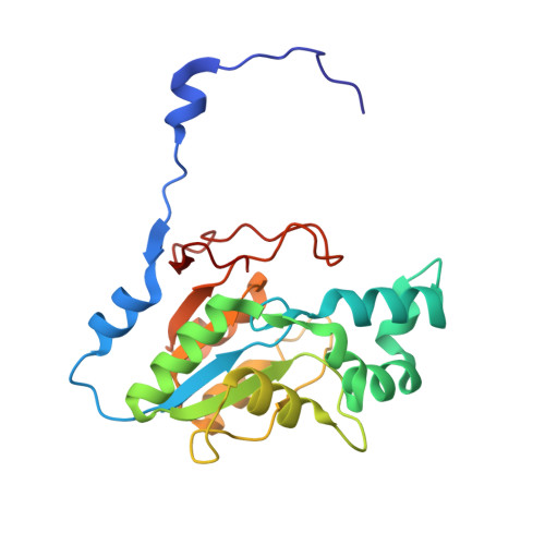
    The Escherichia coli gene aphA codes for a periplasmic acid phosphatase called AphA, belonging to class B bacterial phosphatases, which is part of the DDDD superfamily of phosphohydrolases. After our first report about its crystal structure, we have started a series of crystallographic studies aimed at understanding of the catalytic mechanism of the enzyme. Here, we report three crystal structures of the AphA enzyme in complex with the hydrolysis products of nucleoside monophosphate substrates and a fourth with a proposed intermediate analogue that appears to be covalently bound to the enzyme. Comparison with the native enzyme structure and with the available X-ray structures of different phosphatases provides clues about the enzyme chemistry and allows us to propose a catalytic mechanism for AphA, and to discuss it with respect to the mechanism of other bacterial and human phosphatases.


  • Organizational Affiliation
    • Dipartimento di Chimica, Università di Siena, Via Aldo Moro, I-53100 Siena, Italy.

Macromolecule Content 

  • Total Structure Weight: 47.94 kDa 
  • Atom Count: 3,565 
  • Modeled Residue Count: 422 
  • Deposited Residue Count: 422 
  • Unique protein chains: 1

Macromolecules

Find similar proteins by:|  3D Structure
Entity ID: 1
MoleculeChains  Sequence LengthOrganismDetailsImage
class B acid phosphatase
A, B
211Escherichia coliMutation(s): 0 
Gene Names: aphAnapA
EC: 3.1.3.2
UniProt
Find proteins for P0AE22 (Escherichia coli (strain K12))
Explore P0AE22 
Go to UniProtKB:  P0AE22
Entity Groups
Sequence Clusters30% Identity50% Identity70% Identity90% Identity95% Identity100% Identity
UniProt GroupP0AE22
Sequence Annotations
Expand
Reference Sequence

Small Molecules

Ligands 6 Unique
IDChains Name / Formula / InChI Key2D Diagram3D Interactions
ADN

Query on ADN


Download:Ideal Coordinates CCD File
I [auth B]ADENOSINE
C10 H13 N5 O4
OIRDTQYFTABQOQ-KQYNXXCUSA-N
SPM

Query on SPM


Download:Ideal Coordinates CCD File
D [auth A]SPERMINE
C10 H26 N4
PFNFFQXMRSDOHW-UHFFFAOYSA-N
AU3

Query on AU3


Download:Ideal Coordinates CCD File
G [auth A]GOLD 3+ ION
Au
CBMIPXHVOVTTTL-UHFFFAOYSA-N
AU

Query on AU


Download:Ideal Coordinates CCD File
F [auth A]GOLD ION
Au
ZBKIUFWVEIBQRT-UHFFFAOYSA-N
PO4

Query on PO4


Download:Ideal Coordinates CCD File
E [auth A]PHOSPHATE ION
O4 P
NBIIXXVUZAFLBC-UHFFFAOYSA-K
MG

Query on MG


Download:Ideal Coordinates CCD File
C [auth A],
H [auth B]
MAGNESIUM ION
Mg
JLVVSXFLKOJNIY-UHFFFAOYSA-N

Experimental Data & Validation

Experimental Data

  • Method: X-RAY DIFFRACTION
  • Resolution: 2.03 Å
  • R-Value Free:  0.223 (Depositor), 0.222 (DCC) 
  • R-Value Work:  0.171 (Depositor) 
  • R-Value Observed: 0.175 (Depositor) 
Space Group: C 1 2 1
Unit Cell:
Length ( Å )Angle ( ˚ )
a = 89.966α = 90
b = 66.544β = 124.02
c = 91.544γ = 90
Software Package:
Software NamePurpose
MOSFLMdata reduction
SCALAdata scaling
MOLREPphasing
REFMACrefinement
CCP4data scaling

Structure Validation

View Full Validation Report


Ligand Structure Quality Assessment 


Entry History 

Deposition Data

Revision History  (Full details and data files)

  • Version 1.0: 2005-11-29
    Type: Initial release
  • Version 1.1: 2008-05-01
    Changes: Version format compliance
  • Version 1.2: 2011-07-13
    Changes: Derived calculations, Version format compliance
  • Version 1.3: 2023-08-23
    Changes: Data collection, Database references, Derived calculations, Refinement description