2B4F | pdb_00002b4f

Structure Of A Cold-Adapted Family 8 Xylanase in complex with substrate


Experimental Data Snapshot

  • Method: X-RAY DIFFRACTION
  • Resolution: 1.95 Å
  • R-Value Free: 
    0.178 (Depositor), 0.147 (DCC) 
  • R-Value Work: 
    0.146 (Depositor), 0.153 (DCC) 
  • R-Value Observed: 
    0.148 (Depositor) 

Starting Model: experimental
View more details

wwPDB Validation 3D Report Full Report

Validation slider image for 2B4F

This is version 2.3 of the entry. See complete history. 

Literature

Oligosaccharide binding in family 8 glycosidases: crystal structures of active-site mutants of the beta-1,4-xylanase pXyl from Pseudoaltermonas haloplanktis TAH3a in complex with substrate and product.

De Vos, D., Collins, T., Nerinckx, W., Savvides, S.N., Claeyssens, M., Gerday, C., Feller, G., Van Beeumen, J.

(2006) Biochemistry 45: 4797-4807

  • DOI: https://doi.org/10.1021/bi052193e
  • Primary Citation Related Structures: 
    2B4F

  • PubMed Abstract: 

    The structures of inactive mutants D144A and E78Q of the glycoside hydrolase family 8 (GH-8) endo-beta-1,4-d-xylanase (pXyl) from the Antarctic bacterium Pseudoalteromonas haloplanktis TAH3a in complex with its substrate xylopentaose (at 1.95 A resolution) and product xylotriose (at 1.9 A resolution) have been determined by X-ray crystallography. A detailed comparative analysis of these with the apo-enzyme and with other GH-8 structures indicates an induced fit mechanism upon ligand binding whereby a number of conformational changes and, in particular, a repositioning of the proton donor into a more catalytically competent position occurs. This has also allowed for the description of protein-ligand interactions in this enzyme and for the demarcation of subsites -3 to +3. An in-depth analysis of each of these subsites gives an insight into the structure-function relationship of this enzyme and the basis of xylose/glucose discrimination in family 8 glycoside hydrolases. Furthermore, the structure of the -1/+1 subsite spanning complex reveals that the substrate is distorted from its ground state conformation. Indeed, structural analysis and in silico docking studies indicate that substrate hydrolysis in GH-8 members is preceded by a conformational change, away from the substrate ground-state chair conformation, to a pretransition state local minimum (2)S(O) conformation.


  • Organizational Affiliation: 
    • Laboratorium voor Eiwitbiochemie en Eiwitengineering, Ghent University, K. L. Ledeganckstraat 35, B-9000 Gent, Belgium.

Macromolecule Content 

  • Total Structure Weight: 47.34 kDa 
  • Atom Count: 3,803 
  • Modeled Residue Count: 404 
  • Deposited Residue Count: 405 
  • Unique protein chains: 1

Macromolecules

Find similar proteins by:|  3D Structure
Entity ID: 1
MoleculeChains  Sequence LengthOrganismDetailsImage
endo-1,4-beta-xylanase405Pseudoalteromonas haloplanktisMutation(s): 1 
EC: 3.2.1.8 (PDB Primary Data), 3.2.1.4 (UniProt)
UniProt
Find proteins for Q8RJN8 (Pseudoalteromonas haloplanktis)
Explore Q8RJN8 
Go to UniProtKB:  Q8RJN8
Entity Groups
Sequence Clusters30% Identity50% Identity70% Identity90% Identity95% Identity100% Identity
UniProt GroupQ8RJN8
Sequence Annotations
Expand
Reference Sequence

Oligosaccharides

Help  
Entity ID: 2
MoleculeChains Length2D Diagram GlycosylationD Interactions
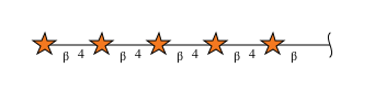
beta-D-xylopyranose-(1-4)-beta-D-xylopyranose-(1-4)-beta-D-xylopyranose-(1-4)-beta-D-xylopyranose-(1-4)-beta-D-xylopyranose
B, C
5N/A
Glycosylation Resources
GlyTouCan: G47101GE
GlyCosmos: G47101GE

Experimental Data & Validation

Experimental Data

  • Method: X-RAY DIFFRACTION
  • Resolution: 1.95 Å
  • R-Value Free:  0.178 (Depositor), 0.147 (DCC) 
  • R-Value Work:  0.146 (Depositor), 0.153 (DCC) 
  • R-Value Observed: 0.148 (Depositor) 
Space Group: P 21 21 21
Unit Cell:
Length ( Å )Angle ( ˚ )
a = 50.711α = 90
b = 91.099β = 90
c = 99.633γ = 90
Software Package:
Software NamePurpose
REFMACrefinement
DENZOdata reduction
SCALEPACKdata scaling
AMoREphasing

Structure Validation

View Full Validation Report


Entry History 

Deposition Data

Revision History  (Full details and data files)

  • Version 1.0: 2006-09-05
    Type: Initial release
  • Version 1.1: 2008-05-01
    Changes: Version format compliance
  • Version 1.2: 2011-07-13
    Changes: Version format compliance
  • Version 1.3: 2015-03-18
    Changes: Derived calculations, Non-polymer description
  • Version 2.0: 2020-07-29
    Type: Remediation
    Reason: Carbohydrate remediation
    Changes: Atomic model, Data collection, Derived calculations, Structure summary
  • Version 2.1: 2021-10-20
    Changes: Database references, Derived calculations, Structure summary
  • Version 2.2: 2023-08-23
    Changes: Data collection, Refinement description
  • Version 2.3: 2024-10-30
    Changes: Structure summary