2B4E | pdb_00002b4e

Crystal Structure of Murine Coronin-1: monoclinic form


Experimental Data Snapshot

  • Method: X-RAY DIFFRACTION
  • Resolution: 2.30 Å
  • R-Value Free: 
    0.229 (Depositor), 0.226 (DCC) 
  • R-Value Work: 
    0.155 (Depositor), 0.156 (DCC) 
  • R-Value Observed: 
    0.159 (Depositor) 

Starting Model: experimental
View more details

wwPDB Validation 3D Report Full Report

Validation slider image for 2B4E

This is version 1.3 of the entry. See complete history

Literature

The crystal structure of murine coronin-1: a regulator of actin cytoskeletal dynamics in lymphocytes

Appleton, B.A.Wu, P.Wiesmann, C.

(2006) Structure 14: 87-96

  • DOI: https://doi.org/10.1016/j.str.2005.09.013
  • Primary Citation Related Structures: 
    2AQ5, 2B4E

  • PubMed Abstract: 

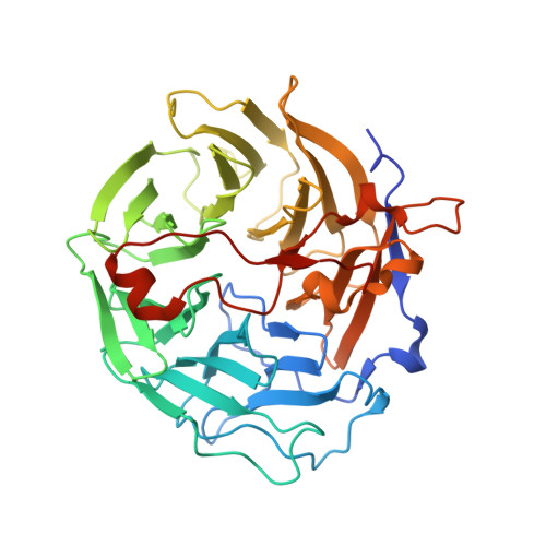
    Mammalian coronin-1 is preferentially expressed in hematopoietic cells and plays a poorly understood role in the dynamic reorganization of the actin cytoskeleton. Sequence analysis of coronin-1 revealed five WD40 repeats that were predicted to form a beta propeller. They are followed by a 130 residue extension and a 30 residue leucine zipper domain that is responsible for multimerization of the protein. Here, we present the crystal structure of murine coronin-1 without the leucine zipper at 1.75 A resolution. Coronin-1 forms a seven-bladed beta propeller composed of the five predicted WD40 repeats and two additional blades that lack any homology to the canonical WD40 motif. The C-terminal extension adopts an extended conformation, packs tightly against the bottom surface of the propeller, and is likely to be required for the structural stability of the propeller. Analysis of charged and conserved surface residues delineate possible binding sites for F-actin on the beta propeller.


  • Organizational Affiliation
    • Department of Protein Engineering, Genentech, Inc., South San Francisco, California 94080, USA.

Macromolecule Content 

  • Total Structure Weight: 44.36 kDa 
  • Atom Count: 3,262 
  • Modeled Residue Count: 386 
  • Deposited Residue Count: 402 
  • Unique protein chains: 1

Macromolecules

Find similar proteins by:|  3D Structure
Entity ID: 1
MoleculeChains  Sequence LengthOrganismDetailsImage
Coronin-1A402Mus musculusMutation(s): 0 
Gene Names: coro1coro1a
UniProt & NIH Common Fund Data Resources
Find proteins for O89053 (Mus musculus)
Explore O89053 
Go to UniProtKB:  O89053
IMPC:  MGI:1345961
Entity Groups
Sequence Clusters30% Identity50% Identity70% Identity90% Identity95% Identity100% Identity
UniProt GroupO89053
Sequence Annotations
Expand
Reference Sequence

Experimental Data & Validation

Experimental Data

  • Method: X-RAY DIFFRACTION
  • Resolution: 2.30 Å
  • R-Value Free:  0.229 (Depositor), 0.226 (DCC) 
  • R-Value Work:  0.155 (Depositor), 0.156 (DCC) 
  • R-Value Observed: 0.159 (Depositor) 
Space Group: P 1 21 1
Unit Cell:
Length ( Å )Angle ( ˚ )
a = 51.358α = 90
b = 80.284β = 111.92
c = 52.889γ = 90
Software Package:
Software NamePurpose
REFMACrefinement
MOSFLMdata reduction
CCP4data scaling
AMoREphasing

Structure Validation

View Full Validation Report


Entry History 

Deposition Data

Revision History  (Full details and data files)

  • Version 1.0: 2005-12-06
    Type: Initial release
  • Version 1.1: 2008-05-01
    Changes: Version format compliance
  • Version 1.2: 2011-07-13
    Changes: Advisory, Version format compliance
  • Version 1.3: 2023-08-23
    Changes: Data collection, Database references, Refinement description