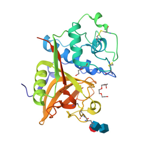
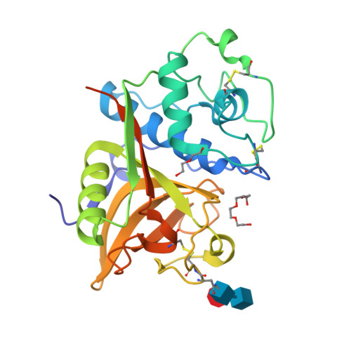
2B1M

Crystal structure of a papain-fold protein without the catalytic cysteine from seeds of Pachyrhizus erosus


Experimental Data Snapshot

  • Method: X-RAY DIFFRACTION
  • Resolution: 2.00 Å
  • R-Value Free: 0.209 
  • R-Value Work: 0.180 

wwPDB Validation   3D Report Full Report


This is version 2.0 of the entry. See complete history


Literature

Crystal structure of a papain-fold protein without the catalytic residue: a novel member in the cysteine proteinase family

Zhang, M.Wei, Z.Chang, S.Teng, M.Gong, W.

(2006) J Mol Biol 358: 97-105

  • DOI: https://doi.org/10.1016/j.jmb.2006.01.065
  • Primary Citation of Related Structures:  
    2B1M, 2B1N

  • PubMed Abstract: 

    A 31kDa cysteine protease, SPE31, was isolated from the seeds of a legume plant, Pachyrizhus erosus. The protein was purified, crystallized and the 3D structure solved using molecular replacement. The cDNA was obtained by RT PCR followed by amplification using mRNA isolated from the seeds of the legume plant as a template. Analysis of the cDNA sequence and the 3D structure indicated the protein to belong to the papain family. Detailed analysis of the structure revealed an unusual replacement of the conserved catalytic Cys with Gly. Replacement of another conserved residue Ala/Gly by a Phe sterically blocks the access of the substrate to the active site. A polyethyleneglycol molecule and a natural peptide fragment were bound to the surface of the active site. Asn159 was found to be glycosylated. The SPE31 cDNA sequence shares several features with P34, a protein found in soybeans, that is implicated in plant defense mechanisms as an elicitor receptor binding to syringolide. P34 has also been shown to interact with vegetative storage proteins and NADH-dependent hydroxypyruvate reductase. These roles suggest that SPE31 and P34 form a unique subfamily within the papain family. The crystal structure of SPE31 complexed with a natural peptide ligand reveals a unique active site architecture. In addition, the clear evidence of glycosylated Asn159 provides useful information towards understanding the functional mechanism of SPE31/P34.


  • Organizational Affiliation

    National Laboratory of Biomacromolecules, Institute of Biophysics, Chinese Academy of Sciences, Beijing 100101, P.R. China.


Macromolecules
Find similar proteins by:  (by identity cutoff)  |  3D Structure
Entity ID: 1
MoleculeChains Sequence LengthOrganismDetailsImage
SPE31246Pachyrhizus erosusMutation(s): 0 
UniProt
Find proteins for Q3Y6U7 (Pachyrhizus erosus)
Explore Q3Y6U7 
Go to UniProtKB:  Q3Y6U7
Entity Groups  
Sequence Clusters30% Identity50% Identity70% Identity90% Identity95% Identity100% Identity
UniProt GroupQ3Y6U7
Glycosylation
Glycosylation Sites: 1
Sequence Annotations
Expand
  • Reference Sequence
Oligosaccharides

Help

Entity ID: 2
MoleculeChains Length2D Diagram Glycosylation3D Interactions
alpha-L-fucopyranose-(1-3)-[2-acetamido-2-deoxy-beta-D-glucopyranose-(1-4)]2-acetamido-2-deoxy-beta-D-glucopyranose
B
3N-Glycosylation
Glycosylation Resources
GlyTouCan:  G11971MR
GlyCosmos:  G11971MR
GlyGen:  G11971MR
Small Molecules
Ligands 2 Unique
IDChains Name / Formula / InChI Key2D Diagram3D Interactions
PG4
Query on PG4

Download Ideal Coordinates CCD File 
C [auth A]TETRAETHYLENE GLYCOL
C8 H18 O5
UWHCKJMYHZGTIT-UHFFFAOYSA-N
PEG
Query on PEG

Download Ideal Coordinates CCD File 
D [auth A]DI(HYDROXYETHYL)ETHER
C4 H10 O3
MTHSVFCYNBDYFN-UHFFFAOYSA-N
Experimental Data & Validation

Experimental Data

  • Method: X-RAY DIFFRACTION
  • Resolution: 2.00 Å
  • R-Value Free: 0.209 
  • R-Value Work: 0.180 
  • Space Group: P 41 21 2
Unit Cell:
Length ( Å )Angle ( ˚ )
a = 61.518α = 90
b = 61.518β = 90
c = 142.191γ = 90
Software Package:
Software NamePurpose
DENZOdata reduction
SCALEPACKdata scaling
CNSrefinement
PDB_EXTRACTdata extraction
AMoREphasing

Structure Validation

View Full Validation Report


Entry History 

Deposition Data

Revision History  (Full details and data files)

  • Version 1.0: 2006-10-03
    Type: Initial release
  • Version 1.1: 2008-05-01
    Changes: Version format compliance
  • Version 1.2: 2011-07-13
    Changes: Non-polymer description, Version format compliance
  • Version 1.3: 2017-10-11
    Changes: Refinement description
  • Version 2.0: 2020-07-29
    Type: Remediation
    Reason: Carbohydrate remediation
    Changes: Atomic model, Data collection, Derived calculations, Structure summary