2B1H | pdb_00002b1h

Crystal structure analysis of anti-HIV-1 V3 Fab 2219 in complex with UG29 peptide


Experimental Data Snapshot

  • Method: X-RAY DIFFRACTION
  • Resolution: 2.00 Å
  • R-Value Free: 
    0.238 (Depositor), 0.243 (DCC) 
  • R-Value Work: 
    0.205 (Depositor), 0.213 (DCC) 
  • R-Value Observed: 
    0.206 (Depositor) 

Starting Model: experimental
View more details

wwPDB Validation 3D Report Full Report

Validation slider image for 2B1H

This is version 1.5 of the entry. See complete history

Literature

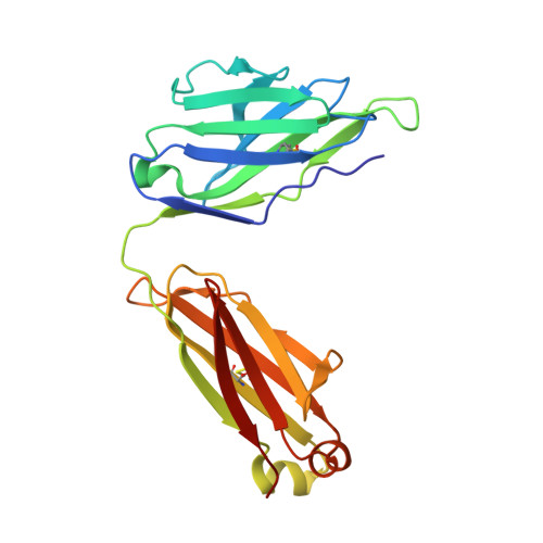
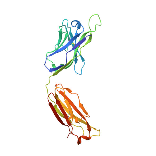
Crystal structures of human immunodeficiency virus type 1 (HIV-1) neutralizing antibody 2219 in complex with three different V3 peptides reveal a new binding mode for HIV-1 cross-reactivity.

Stanfield, R.L.Gorny, M.K.Zolla-Pazner, S.Wilson, I.A.

(2006) J Virol 80: 6093-6105

  • DOI: https://doi.org/10.1128/JVI.00205-06
  • Primary Citation Related Structures: 
    2B0S, 2B1A, 2B1H

  • PubMed Abstract: 

    Human monoclonal antibody 2219 is a neutralizing antibody isolated from a human immunodeficiency virus type 1-infected individual. 2219 was originally selected for binding to a V3 fusion protein and can neutralize primary isolates from subtypes B, A, and F. Thus, 2219 represents a cross-reactive, human anti-V3 antibody. Fab 2219 binds to one face of the variable V3 beta-hairpin, primarily contacting conserved residues on the N-terminal beta-strand of V3, leaving the V3 crown or tip largely accessible. Three V3/2219 complexes reveal the antibody-bound conformations for both the N- and C-terminal regions that flank the V3 crown and illustrate how twisting of the V3 loop alters the relative dispositions and pairing of the amino acids in the adjacent V3 beta-strands and how the antibody can accommodate V3 loops with different sequences.


  • Organizational Affiliation
    • Department of Molecular Biology, The Scripps Research Institute, La Jolla, CA 92037, USA. robyn@scripps.edu

Macromolecule Content 

  • Total Structure Weight: 49.7 kDa 
  • Atom Count: 3,572 
  • Modeled Residue Count: 457 
  • Deposited Residue Count: 464 
  • Unique protein chains: 3

Macromolecules

Find similar proteins by:|  3D Structure
Entity ID: 1
MoleculeChains  Sequence LengthOrganismDetailsImage
Fab 2219, light chainA [auth L]215Homo sapiensMutation(s): 0 
Find similar proteins by:|  3D Structure
Entity ID: 2
MoleculeChains  Sequence LengthOrganismDetailsImage
Fab 2219, heavy chainB [auth H]226Homo sapiensMutation(s): 0 
Find similar proteins by:  Sequence   |   3D Structure  
Entity ID: 3
MoleculeChains  Sequence LengthOrganismDetailsImage
UG29 peptide of Exterior membrane glycoprotein GP120C [auth P]23N/AMutation(s): 0 

Experimental Data & Validation

Experimental Data

  • Method: X-RAY DIFFRACTION
  • Resolution: 2.00 Å
  • R-Value Free:  0.238 (Depositor), 0.243 (DCC) 
  • R-Value Work:  0.205 (Depositor), 0.213 (DCC) 
  • R-Value Observed: 0.206 (Depositor) 
Space Group: P 21 21 21
Unit Cell:
Length ( Å )Angle ( ˚ )
a = 62.899α = 90
b = 94.501β = 90
c = 96.617γ = 90
Software Package:
Software NamePurpose
REFMACrefinement
SCALEPACKdata scaling
AMoREphasing
CNSrefinement
DENZOdata reduction

Structure Validation

View Full Validation Report


Entry History 

Deposition Data

Revision History  (Full details and data files)

  • Version 1.0: 2006-07-04
    Type: Initial release
  • Version 1.1: 2008-05-01
    Changes: Version format compliance
  • Version 1.2: 2011-07-13
    Changes: Advisory, Version format compliance
  • Version 1.3: 2019-07-24
    Changes: Data collection, Refinement description
  • Version 1.4: 2023-08-23
    Changes: Data collection, Database references, Refinement description
  • Version 1.5: 2024-11-13
    Changes: Structure summary