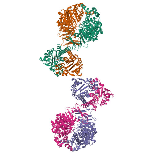
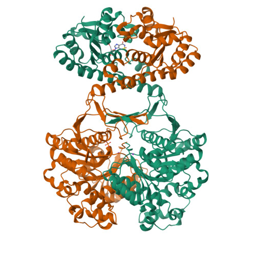
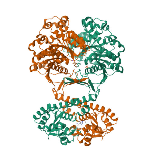
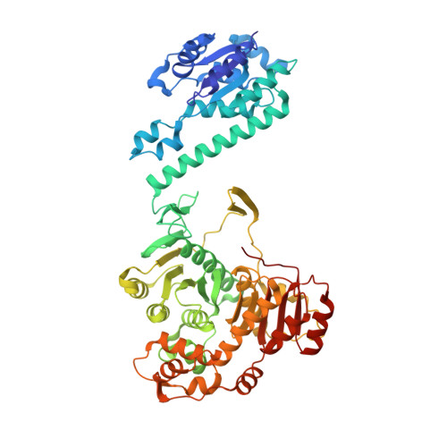
2B1G

Crystal structures of transition state analogue inhibitors of inosine monophosphate cyclohydrolase


Experimental Data Snapshot

  • Method: X-RAY DIFFRACTION
  • Resolution: 2.10 Å
  • R-Value Free: 0.248 
  • R-Value Work: 0.199 
  • R-Value Observed: 0.243 

Starting Model: experimental
View more details

wwPDB Validation   3D Report Full Report


Ligand Structure Quality Assessment 


This is version 1.4 of the entry. See complete history


Literature

Structure-based Design, Synthesis, Evaluation, and Crystal Structures of Transition State Analogue Inhibitors of Inosine Monophosphate Cyclohydrolase.

Xu, L.Chong, Y.Hwang, I.D'Onofrio, A.Amore, K.Beardsley, G.P.Li, C.Olson, A.J.Boger, D.L.Wilson, I.A.

(2007) J Biol Chem 282: 13033-13046

  • DOI: https://doi.org/10.1074/jbc.M607293200
  • Primary Citation of Related Structures:  
    2B1G, 2B1I, 2IU0, 2IU3

  • PubMed Abstract: 

    The inosine monophosphate cyclohydrolase (IMPCH) component (residues 1-199) of the bifunctional enzyme aminoimidazole-4-carboxamide ribonucleotide transformylase (AICAR Tfase, residues 200-593)/IMPCH (ATIC) catalyzes the final step in the de novo purine biosynthesis pathway that produces IMP. As a potential target for antineoplastic intervention, we designed IMPCH inhibitors, 1,5-dihydroimidazo[4,5-c][1,2,6]thiadiazin-4(3H)-one 2,2-dioxide (heterocycle, 1), the corresponding nucleoside (2), and the nucleoside monophosphate (nucleotide) (3), as mimics of the tetrahedral intermediate in the cyclization reaction. All compounds are competitive inhibitors against IMPCH (K(i) values = 0.13-0.23 microm) with the simple heterocycle 1 exhibiting the most potent inhibition (K(i) = 0.13 microm). Crystal structures of bifunctional ATIC in complex with nucleoside 2 and nucleotide 3 revealed IMPCH binding modes similar to that of the IMPCH feedback inhibitor, xanthosine 5'-monophosphate. Surprisingly, the simpler heterocycle 1 had a completely different IMPCH binding mode and was relocated to the phosphate binding pocket that was identified from previous xanthosine 5'-monophosphate structures. The aromatic imidazole ring interacts with a helix dipole, similar to the interaction with the phosphate moiety of 3. The crystal structures not only revealed the mechanism of inhibition of these compounds, but they now serve as a platform for future inhibitor improvements. Importantly, the nucleoside-complexed structure supports the notion that inhibitors lacking a negatively charged phosphate can still inhibit IMPCH activity with comparable potency to phosphate-containing inhibitors. Provocatively, the nucleotide inhibitor 3 also binds to the AICAR Tfase domain of ATIC, which now provides a lead compound for the design of inhibitors that simultaneously target both active sites of this bifunctional enzyme.


  • Organizational Affiliation

    Department of Molecular Biology, The Scripps Research Institute, La Jolla, California 92037, USA.


Macromolecules
Find similar proteins by:  (by identity cutoff)  |  3D Structure
Entity ID: 1
MoleculeChains Sequence LengthOrganismDetailsImage
Bifunctional purine biosynthesis protein PURH
A, B, C, D
593Gallus gallusMutation(s): 0 
Gene Names: ATICPURH
EC: 3.5.4.10 (PDB Primary Data), 2.1.2.3 (PDB Primary Data)
UniProt
Find proteins for P31335 (Gallus gallus)
Explore P31335 
Go to UniProtKB:  P31335
Entity Groups  
Sequence Clusters30% Identity50% Identity70% Identity90% Identity95% Identity100% Identity
UniProt GroupP31335
Sequence Annotations
Expand
  • Reference Sequence
Small Molecules
Ligands 3 Unique
IDChains Name / Formula / InChI Key2D Diagram3D Interactions
13A
Query on 13A

Download Ideal Coordinates CCD File 
G [auth A],
L [auth D]
7-(3,4-DIHYDROXY-5R-HYDROXYMETHYLTETRAHYDROFURAN-2-YL)-2,2-DIOXO-1,2R,3R,7-TETRAHYDRO-2L6-IMIDAZO[4,5-C][1,2,6]THIADIAZIN-4S-ONE
C9 H12 N4 O7 S
IHLOTZVBEUFDMD-UUOKFMHZSA-N
PO4
Query on PO4

Download Ideal Coordinates CCD File 
E [auth A],
I [auth C]
PHOSPHATE ION
O4 P
NBIIXXVUZAFLBC-UHFFFAOYSA-K
K
Query on K

Download Ideal Coordinates CCD File 
F [auth A],
H [auth B],
J [auth C],
K [auth D]
POTASSIUM ION
K
NPYPAHLBTDXSSS-UHFFFAOYSA-N
Binding Affinity Annotations 
IDSourceBinding Affinity
13A PDBBind:  2B1G Ki: 230 (nM) from 1 assay(s)
Experimental Data & Validation

Experimental Data

  • Method: X-RAY DIFFRACTION
  • Resolution: 2.10 Å
  • R-Value Free: 0.248 
  • R-Value Work: 0.199 
  • R-Value Observed: 0.243 
  • Space Group: P 1
Unit Cell:
Length ( Å )Angle ( ˚ )
a = 57.2α = 80.8
b = 62β = 81.5
c = 196.6γ = 90
Software Package:
Software NamePurpose
DENZOdata reduction
SCALEPACKdata scaling
AMoREphasing
REFMACrefinement
PDB_EXTRACTdata extraction
HKL-2000data reduction

Structure Validation

View Full Validation Report


Ligand Structure Quality Assessment 


Entry History 

Deposition Data

Revision History  (Full details and data files)

  • Version 1.0: 2006-11-21
    Type: Initial release
  • Version 1.1: 2008-04-01
    Changes: Version format compliance
  • Version 1.2: 2011-07-13
    Changes: Derived calculations, Version format compliance
  • Version 1.3: 2017-10-11
    Changes: Refinement description
  • Version 1.4: 2023-08-23
    Changes: Data collection, Database references, Derived calculations, Refinement description