2AZ1

Structure of a halophilic nucleoside diphosphate kinase from Halobacterium salinarum


Experimental Data Snapshot

  • Method: X-RAY DIFFRACTION
  • Resolution: 2.35 Å
  • R-Value Free: 0.289 
  • R-Value Work: 0.217 
  • R-Value Observed: 0.220 

Starting Model: experimental
View more details

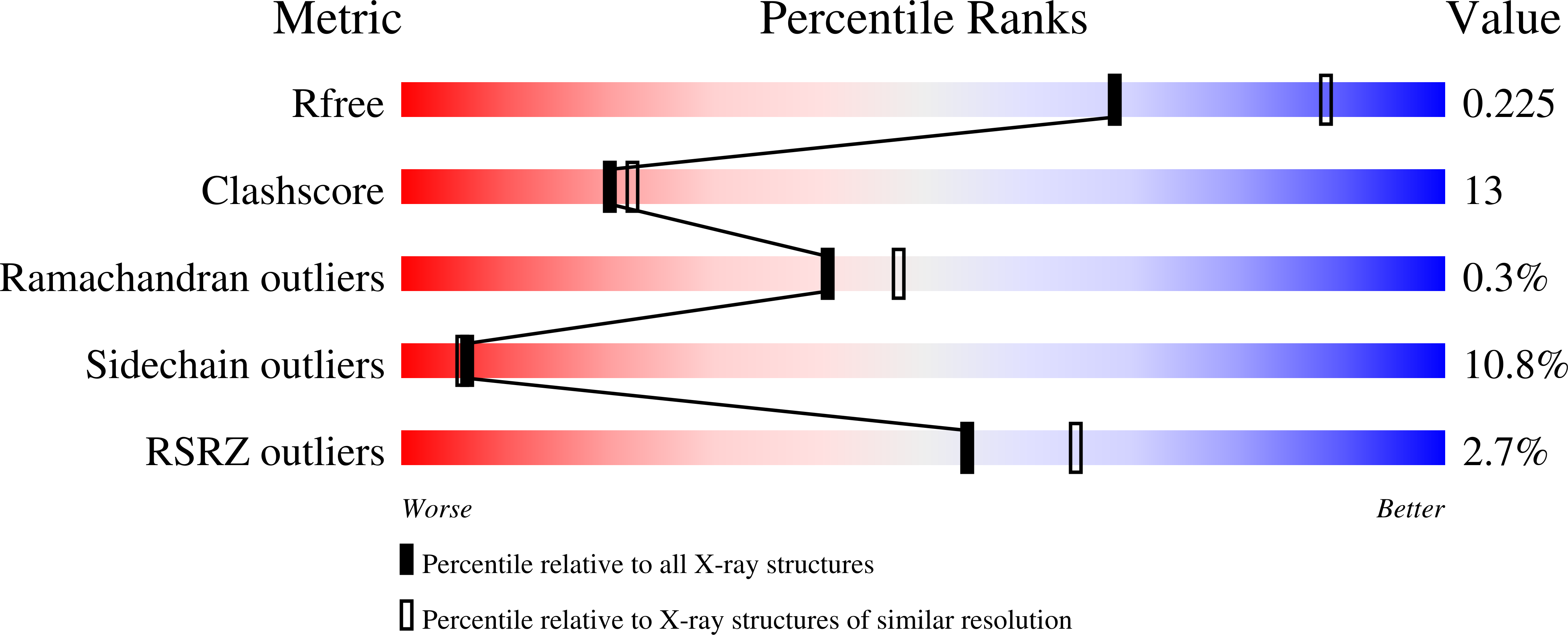
wwPDB Validation   3D Report Full Report


This is version 1.4 of the entry. See complete history


Literature

Structure of a halophilic nucleoside diphosphate kinase from Halobacterium salinarum

Besir, H.Zeth, K.Bracher, A.Heider, U.Ishibashi, M.Tokunaga, M.Oesterhelt, D.

(2005) FEBS Lett 579: 6595-6600

  • DOI: https://doi.org/10.1016/j.febslet.2005.10.052
  • Primary Citation of Related Structures:  
    2AZ1, 2AZ3

  • PubMed Abstract: 

    Nucleoside diphosphate kinase from the halophilic archaeon Halobacterium salinarum was crystallized in a free state and a substrate-bound form with CDP. The structures were solved to a resolution of 2.35 and 2.2A, respectively. Crystals with the apo-form were obtained with His6-tagged enzyme, whereas the untagged form was used for co-crystallization with the nucleotide. Crosslinking under different salt and pH conditions revealed a stronger oligomerization tendency for the tagged protein at low and high salt concentrations. The influence of the His6-tag on the halophilic nature of the enzyme is discussed on the basis of the observed structural properties.


  • Organizational Affiliation

    Max-Planck-Institut für Biochemie, Abteilung Membranbiochemie, Am Klopferspitz 18, 82152 Martinsried, Germany. hbesir@biochem.mpg.de


Macromolecules
Find similar proteins by:  (by identity cutoff)  |  3D Structure
Entity ID: 1
MoleculeChains Sequence LengthOrganismDetailsImage
Nucleoside diphosphate kinase
A, B, C, D, E
A, B, C, D, E, F
181Halobacterium salinarumMutation(s): 0 
EC: 2.7.4.6
UniProt
Find proteins for P61136 (Halobacterium salinarum (strain ATCC 700922 / JCM 11081 / NRC-1))
Explore P61136 
Go to UniProtKB:  P61136
Entity Groups  
Sequence Clusters30% Identity50% Identity70% Identity90% Identity95% Identity100% Identity
UniProt GroupP61136
Sequence Annotations
Expand
  • Reference Sequence
Experimental Data & Validation

Experimental Data

  • Method: X-RAY DIFFRACTION
  • Resolution: 2.35 Å
  • R-Value Free: 0.289 
  • R-Value Work: 0.217 
  • R-Value Observed: 0.220 
  • Space Group: P 21 21 21
Unit Cell:
Length ( Å )Angle ( ˚ )
a = 69.28α = 90
b = 108.7β = 90
c = 119.65γ = 90
Software Package:
Software NamePurpose
REFMACrefinement
MAR345data collection
XDSdata scaling
MOLREPphasing

Structure Validation

View Full Validation Report


Entry History 

Deposition Data

Revision History  (Full details and data files)

  • Version 1.0: 2005-12-20
    Type: Initial release
  • Version 1.1: 2008-05-01
    Changes: Version format compliance
  • Version 1.2: 2011-07-13
    Changes: Version format compliance
  • Version 1.3: 2017-10-11
    Changes: Refinement description
  • Version 1.4: 2023-10-25
    Changes: Data collection, Database references, Derived calculations, Refinement description