2AWC | pdb_00002awc

deoxy-DcrH-Hr


Experimental Data Snapshot

  • Method: X-RAY DIFFRACTION
  • Resolution: 2.20 Å
  • R-Value Free: 
    0.253 (Depositor), 0.251 (DCC) 
  • R-Value Work: 
    0.204 (Depositor), 0.199 (DCC) 
  • R-Value Observed: 
    0.204 (Depositor) 

wwPDB Validation 3D Report Full Report

Validation slider image for 2AWC

This is version 1.3 of the entry. See complete history

Literature

Structural Basis for O(2) Sensing by the Hemerythrin-like Domain of a Bacterial Chemotaxis Protein: Substrate Tunnel and Fluxional N Terminus

Isaza, C.E.Silaghi-Dumitrescu, R.Iyer, R.B.Kurtz, D.M.Chan, M.K.

(2006) Biochemistry 45: 9023-9031

  • DOI: https://doi.org/10.1021/bi0607812
  • Primary Citation Related Structures: 
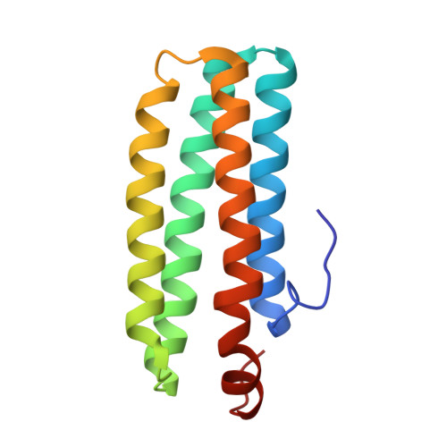
    2AVK, 2AWC, 2AWY

  • PubMed Abstract: 

    The methyl-accepting chemotaxis protein, DcrH, from the anaerobic sulfate-reducing bacterium, Desulfovibrio vulgaris (Hildenborough), has a hemerythrin-like domain, DcrH-Hr, at its C terminus. DcrH-Hr was previously shown to contain a diiron site that binds O2, suggesting an O2-sensing function. X-ray crystal structures of diferric (met-), azido-diferric (azidomet-), and diferrous (deoxy-) DcrH-Hr reveal a "substrate tunnel" distinct from that in invertebrate hemerythrins. This tunnel is proposed to facilitate the rapid autoxidation of oxy-DcrH-Hr and suggests that sensing is triggered by O2 binding and subsequent oxidation of the diferrous active site. The N-terminal loop of DcrH-Hr is highly ordered in both met- and azidomet-DcrH-Hr but is disordered in deoxy-DcrH-Hr. These redox-dependent conformational differences presumably transduce the sensory signal of DcrH-Hr to the neighboring methylation domain in the full-length receptor. Given the putative cytoplasmic localization of its Hr-like O2-sensing domain, DcrH is proposed to serve a role in negative aerotaxis (anaerotaxis).


  • Organizational Affiliation
    • The Ohio State Biophysics Program, The Ohio State University, 484 West 12th Avenue, Columbus, Ohio 43210, USA.

Macromolecule Content 

  • Total Structure Weight: 15.72 kDa 
  • Atom Count: 1,232 
  • Modeled Residue Count: 130 
  • Deposited Residue Count: 130 
  • Unique protein chains: 1

Macromolecules

Find similar proteins by:|  3D Structure
Entity ID: 1
MoleculeChains  Sequence LengthOrganismDetailsImage
hemerythrin-like domain protein DcrH130Nitratidesulfovibrio vulgarisMutation(s): 0 
UniProt
Find proteins for Q9REU3 (Nitratidesulfovibrio vulgaris)
Explore Q9REU3 
Go to UniProtKB:  Q9REU3
Entity Groups
Sequence Clusters30% Identity50% Identity70% Identity90% Identity95% Identity100% Identity
UniProt GroupQ9REU3
Sequence Annotations
Expand
Reference Sequence

Small Molecules

Ligands 1 Unique
IDChains Name / Formula / InChI Key2D Diagram3D Interactions
FEO

Query on FEO


Download:Ideal Coordinates CCD File
B [auth A]MU-OXO-DIIRON
Fe2 O
NPMYUMBHPJGBFA-UHFFFAOYSA-N

Experimental Data & Validation

Experimental Data

  • Method: X-RAY DIFFRACTION
  • Resolution: 2.20 Å
  • R-Value Free:  0.253 (Depositor), 0.251 (DCC) 
  • R-Value Work:  0.204 (Depositor), 0.199 (DCC) 
  • R-Value Observed: 0.204 (Depositor) 
Space Group: P 1 21 1
Unit Cell:
Length ( Å )Angle ( ˚ )
a = 43.98α = 90
b = 33.12β = 96.02
c = 44.7γ = 90
Software Package:
Software NamePurpose
CNSrefinement
Blu-Icedata collection
MOSFLMdata reduction
PHASERphasing

Structure Validation

View Full Validation Report


Entry History 

Deposition Data

Revision History  (Full details and data files)

  • Version 1.0: 2006-08-15
    Type: Initial release
  • Version 1.1: 2008-05-01
    Changes: Version format compliance
  • Version 1.2: 2011-07-13
    Changes: Version format compliance
  • Version 1.3: 2024-02-14
    Changes: Data collection, Database references, Derived calculations