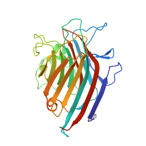
2AUY

Pterocarpus angolensis lectin in complex with the trisaccharide GlcNAc(b1-2)Man(a1-3)Man


Experimental Data Snapshot

  • Method: X-RAY DIFFRACTION
  • Resolution: 1.95 Å
  • R-Value Free: 0.193 
  • R-Value Work: 0.175 
  • R-Value Observed: 0.177 

Starting Model: experimental
View more details

wwPDB Validation   3D Report Full Report


This is version 3.1 of the entry. See complete history


Literature

Structural basis for the recognition of complex-type biantennary oligosaccharides by Pterocarpus angolensis lectin.

Buts, L.Garcia-Pino, A.Imberty, A.Amiot, N.Boons, G.-J.Beeckmans, S.Versees, W.Wyns, L.Loris, R.

(2006) FEBS J 273: 2407-2420

  • DOI: https://doi.org/10.1111/j.1742-4658.2006.05248.x
  • Primary Citation of Related Structures:  
    2AR6, 2ARB, 2ARE, 2ARX, 2AUY

  • PubMed Abstract: 

    The crystal structure of Pterocarpus angolensis lectin is determined in its ligand-free state, in complex with the fucosylated biantennary complex type decasaccharide NA2F, and in complex with a series of smaller oligosaccharide constituents of NA2F. These results together with thermodynamic binding data indicate that the complete oligosaccharide binding site of the lectin consists of five subsites allowing the specific recognition of the pentasaccharide GlcNAc beta(1-2)Man alpha(1-3)[GlcNAc beta(1-2)Man alpha(1-6)]Man. The mannose on the 1-6 arm occupies the monosaccharide binding site while the GlcNAc residue on this arm occupies a subsite that is almost identical to that of concanavalin A (con A). The core mannose and the GlcNAc beta(1-2)Man moiety on the 1-3 arm on the other hand occupy a series of subsites distinct from those of con A.


  • Organizational Affiliation

    Laboratorium voor Ultrastructuur, Vrije Universiteit Brussel and Department of Molecular and Cellular Interactions, Vlaams Interuniversitair Instituut voor Biotechnologie, Belgium.


Macromolecules
Find similar proteins by:  (by identity cutoff)  |  3D Structure
Entity ID: 1
MoleculeChains Sequence LengthOrganismDetailsImage
lectin
A, B
252Pterocarpus angolensisMutation(s): 0 
UniProt
Find proteins for Q8GSD2 (Pterocarpus angolensis)
Explore Q8GSD2 
Go to UniProtKB:  Q8GSD2
Entity Groups  
Sequence Clusters30% Identity50% Identity70% Identity90% Identity95% Identity100% Identity
UniProt GroupQ8GSD2
Sequence Annotations
Expand
  • Reference Sequence
Oligosaccharides

Help

Entity ID: 2
MoleculeChains Length2D Diagram Glycosylation3D Interactions
2-acetamido-2-deoxy-beta-D-glucopyranose-(1-2)-alpha-D-mannopyranose-(1-3)-methyl alpha-D-mannopyranoside
C, D
3N/A
Glycosylation Resources
GlyTouCan:  G62814YX
GlyCosmos:  G62814YX
Experimental Data & Validation

Experimental Data

  • Method: X-RAY DIFFRACTION
  • Resolution: 1.95 Å
  • R-Value Free: 0.193 
  • R-Value Work: 0.175 
  • R-Value Observed: 0.177 
  • Space Group: P 21 21 21
Unit Cell:
Length ( Å )Angle ( ˚ )
a = 56.8α = 90
b = 83.14β = 90
c = 123.02γ = 90
Software Package:
Software NamePurpose
DENZOdata reduction
SCALEPACKdata scaling
AMoREphasing
CNSrefinement

Structure Validation

View Full Validation Report


Entry History 

Deposition Data

Revision History  (Full details and data files)

  • Version 1.0: 2006-08-08
    Type: Initial release
  • Version 1.1: 2008-05-01
    Changes: Version format compliance
  • Version 1.2: 2011-07-13
    Changes: Non-polymer description, Version format compliance
  • Version 1.3: 2017-10-11
    Changes: Refinement description
  • Version 2.0: 2019-12-25
    Changes: Data collection, Database references, Derived calculations, Polymer sequence
  • Version 3.0: 2020-07-29
    Type: Remediation
    Reason: Carbohydrate remediation
    Changes: Atomic model, Data collection, Derived calculations, Structure summary
  • Version 3.1: 2023-08-23
    Changes: Data collection, Database references, Derived calculations, Refinement description, Structure summary